Fear
A New Path for Dealing With Fear
A new study suggests new paths for treating anxiety that bypass exposure.
Posted January 30, 2021 Reviewed by Gary Drevitch

Society has attached a negative connotation to the emotion of fear—and for valid reasons. The last time you felt fear may have been in preparation for a job interview, or reviewing for an entrance exam, or dealing with the concerns associated with the pandemic. Indeed, most people would characterize these as instances related to some form of fear.
Thus, fear is generally recognized as an unpleasant emotion, and people often try to avoid it. However, from an evolutionary perspective, it actually serves a vital role in keeping you safe from potential danger (Öhman, 2000). For example, one may have heard of the so-called “fight-or-flight” response, which is one such occurrence of your body responding to fear or perceived threat. In this response, your body releases hormones that ultimately result in an increase in heart rate and blood pressure, preparing your body to spring into action (hence, “fight-or-flight”) (Gordan et al., 2015).
Nevertheless, society’s generally negative perception of fear still deserves attention, since heightened fear is a significant precursor to various anxiety-related disorders (Rothbaum & David, 2003). In fact, fear-induced disorders are some of the most common psychiatric illnesses, amassing to hundreds of millions of individuals worldwide (T. Vos et al., 2016). Thus, despite fear being a natural—and even necessary—physiological response to stimuli, there is a need to optimize treatments for fear mitigation when fear is excessive and causes psychological distress.
Exposure therapy is known to be a highly effective psychological treatment for anxiety and post-traumatic disorders. People who suffer from these disorders tend to avoid the anxiety-provoking situation. For example, people with social anxiety will refrain from public speaking. This avoidance momentarily reduces the anxiety, but paradoxically increases the degree to which the same event/stimuli causes anxiety next time. Exposure therapy aims to break this vicious cycle. In exposure therapy, psychologists expose individuals to these feared situations, objects, or activities in a gradual manner, thus making them confront their fears and realize there is nothing to be afraid of. For example, someone with a fear of flying might be put in a virtual environment mimicking the sights, sounds, and smells of an airplane (Rothbaum et al., 1997).
However, studies show that exposure therapy, especially with adolescents, faces unique challenges. Exposure therapy relies on the prefrontal regions of the brain, which fully mature in the mid-twenties (Arain et al., 2013). The ethical and physiological difficulties presented by exposure therapy usher in a potential need for alternative treatments. One promising avenue of research comes from the safety signal learning literature. Significantly, this safety learning pathway is different from the neural pathway that supports extinction learning. Thus, there may be opportunities to leverage safety learning to enhance fear reduction when the pathway that supports extinction learning is still developing, and Prof. Dylan Gee from the Yale Psychology department, has offered important insight as to why this may be so.
Safety Signal Learning
What exactly is safety signal learning? As a child, you might have had that one toy or blanket that you constantly kept by your side. Whether consciously or subconsciously, you learned to associate it with comfort and safety. In times of fear or isolation, you would tightly hug that blanket and feel better. This is precisely what safety signal learning is—associating certain objects, sounds, or environmental stimuli with being safe and/or the absence of aversive events.
In their study, Meyer, Odriozola and their colleagues investigate the physiological and neural mechanisms underlying safety signal learning. They confirmed that a neural region potentially involved in safety signal learning is the ventral hippocampus. This brain region is located in the temporal lobe, which is important for learning and memory.
To investigate ventral hippocampal activity, the team performed a series of experiments on both mice and human subjects. Scientists often use mice in their research because mice reproduce quickly and are cost-efficient (Vanhooren & Libert, 2013). Since they also have a very similar genome to the human genome (Vanhooren & Libert, 2013), neural behavior in mice can readily teach us about neural behavior in humans. The researchers used fiber photometry and functional Magnetic Resonance Imaging (fMRI) to monitor neural activity in the mice and human hippocampal regions respectively. Briefly, fiber photometry is an imaging method that tracks neuron activity by locating where there is elevated intracellular calcium levels, which occurs when neurons are active in mice. fMRI is also an imaging method, but instead of tracking calcium levels, it measures brain activity by detecting changes associated with blood flow in the brain.
Meyer & Odriozola’s Experiment and Results
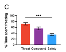
In the first part of their experiment, Meyer and Odriozola et al. trained mice to differentiate between a tone associated with a mild foot shock (i.e., a threat cue; ouch!) and another, distinct tone associated with no threat (i.e., a safety cue). This training process occurred over the course of four days. Then, the researchers entered the testing phase, in which mice were randomly exposed to either the threat cue, the safety cue, or a simultaneous “compound” of both cues (imagine playing one note on the piano; versus playing another, distinct note on the piano; versus playing both notes at the same time). To quantify fear, the researchers measured a species-specific response to fear called freezing time, or the amount of time the mice did not move after hearing a tone (Curzon et al., 2009). Longer freezing time is interpreted as a higher level of anxiety.
They discovered that the time spent freezing was the highest in response to the threat cue, the second-highest in response to the combined cue, and the lowest in response to the safety cue. In other words, they found that a clearer presence of the “safety signal” reduced the physiological response of fear-induced freezing. It is similar to a situation in which a young child goes to a dental exam (scary!). Bringing the child’s beloved blanket or toy—their “safety signal”—may very well help calm their nerves.

To verify that the findings in mice are relevant to humans, the researchers designed a similar task for human participants. Just like the mice experiment, there were similar learning and testing phases to which each participant was exposed. As expected, in parallel with the results of the mice experiment, they found that measured fear was highest in response to the threat cue, second-highest in response to the “compound” cue, and lowest in response to the safety cue.
One question remained: What was the mechanism by which this safety signal learning occurred? Using fiber photometry and fMRI, the researchers found that neural activity was most prominent in the ventral hippocampus, and that this activity was closely linked to the prelimbic prefrontal cortex, another portion of the brain important for a variety of complex cognitive behaviors including planning and personality development. This connection was observed in homologous regions of mice and human brains.
Implications of the Study
These are quite significant results. New insights about safety signal learning could provide clues as to how to optimize treatment for anxiety disorders. The experimental results add to the existing literature surrounding fear reduction and have the potential to optimize treatment for related disorders. These findings can be summarized in three points:
- Safety signal learning may operate on a different timescale than extinction learning, which forms the basis for exposure treatments.
- Safety signal learning engages the ventral hippocampus, a part of the brain involved in learning and emotions.
- Specifically, during this process of safety learning, the ventral hippocampus interacts with the prelimbic prefrontal cortex in mice, and the homologous regions in humans. Significantly, this safety learning pathway is different from the neural pathway that supports extinction learning. Thus, there may be opportunities to leverage safety learning to enhance fear reduction when the pathway that supports extinction learning is still developing. For example, the region of the brain involved in exposure therapy develops more slowly and does not reach full maturity until age 25 (Arain et al., 2013).
From the data collected by Meyer and Odriozola, it is clear that treatments for anxiety disorders are still very much in the process of being optimized. However, given the current direction and attention given to this area of study, we are optimistic about the kinds of treatments that are being developed.
Adam Zhang (Yale undergraduate) and Reuma Gadassi Polack (postdoctoral fellow at Yale) contributed to this post.
LinkedIn image: CGN089/Shutterstock. Facebook image: cheapbooks/Shutterstock
References
Adolphs R. (2013). The biology of fear. Current biology : CB, 23(2), R79–R93. https://doi.org/10.1016/j.cub.2012.11.055
Arain, M., Haque, M., Johal, L., Mathur, P., Nel, W., Rais, A., Sandhu, R., & Sharma, S. (2013). Maturation of the adolescent brain. Neuropsychiatric disease and treatment, 9, 449–461. https://doi.org/10.2147/NDT.S39776
B. O. Rothbaum, M. Davis. (2003). Applying learning principles to the treatment of post- trauma reactions. Ann. N. Y. Acad. Sci. 1008, 112–121
Curzon P, Rustay NR, Browman KE. (2009). Cued and Contextual Fear Conditioning for Rodents. In: Buccafusco JJ, editor. Methods of Behavior Analysis in Neuroscience. 2nd edition. Boca Raton (FL): CRC Press/Taylor & Francis;. Chapter 2. Available from: https://www.ncbi.nlm.nih.gov/books/NBK5223/
Fere, C. (1988). Note on changes in electrical resistance under the effectof sensory stimulation and emotion. Comptes Rendus des Seances de laSociete de Biologie (Series 9), 5, 217-219.
Gola, J. A., Beidas, R. S., Antinoro-Burke, D., Kratz, H. E., & Fingerhut, R. (2016). Ethical Considerations in Exposure Therapy With Children. Cognitive and behavioral practice, 23(2), 184–193. https://doi.org/10.1016/j.cbpra.2015.04.003
Gordan R, Gwathmey JK, Xie LH. (2015). Autonomic and endocrine control of cardiovascular function. World J Cardiol, 7(4):204-14. doi:10.4330/wjc.v7.i4.204
Meyer*, H. C., Odriozola*, P., Cohodes, E. M., Mandell, J. D., Li, A., Yang, R., ... , Lee*, F. S., Gee*, D. G. (2019). Ventral hippocampus interacts with prelimbic cortex during inhibition of threat response via learned safety in both mice and humans. Proceedings of the National Academy of Sciences, 116(52), 26970-26979.
Öhman, A. (2000). "Fear and anxiety: Evolutionary, cognitive, and clinical perspectives". In M. Lewis & J.M. Haviland-Jones (Eds.). Handbook of emotions. pp. 573–93. New York: The Guilford Press.
Rothbaum, B. O., Hodges, L., & Kooper, R. (1997). Virtual reality exposure therapy. Journal of Psychotherapy Practice & Research, 6(3), 219–226.
T. Vos et al. (2016). GBD 2015 Disease and Injury Incidence and Prevalence Collaborators, Global, regional, and national incidence, prevalence, and years lived with disability for 310 diseases and injuries, 1990-2015: A systematic analysis for the Global Burden of Disease Study 2015. Lancet 388, 1545–1602
Vanhooren V, Libert C. (2012) The mouse as a model organism in aging research: usefulness, pitfalls and possibilities. Ageing Res Rev. 2013 Jan;12(1):8-21. doi: 10.1016/j.arr.2012.03.010. Epub. PMID: 22543101.


