Autism
Your Eyes Are a Window Into the Inner Workings of Your Brain
Rapid eye movements rely on multiplex functions of cerebellar Purkinje neurons.
Posted July 28, 2016

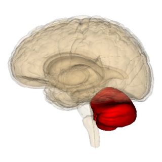
Every morning, I wake up hoping there will be breaking news that helps us better understand the inner workings of the cerebellum (Latin for "little brain"). The cerebellum is primarily known for its role in all types of sensorimotor coordination and creating fluidity of movement. Today, a new study by a team of international researchers reveals more clues as to how specific neurons in the cerebellum are linked to our eyes being a window into the brain.
I know that looking forward to new 'cerebellar' (relating to or located in the cerebellum) research is an extremely esoteric reason to spring out of bed in the morning... But, for me, decoding the mysteries of Purkinje cells and the cerebellum is deeply personal.
Before his sudden death in 2007, my father—who was a neuroscientist, neurosurgeon, and author of The Fabric of Mind (Viking)—dedicated the final years of his life attempting to solve multifaceted and enigmatic riddles of the cerebellum. He died before advances in neuroimaging technology made it possible for him to realize his dreams and hypotheses. At his funeral, I made a vow that I would do my best to keep my dad's passion for "the little brain" alive throughout the twenty-first century.
Over the past decade, I've kept my antennae up for advances in neuroscience technology and strived to connect-the-dots of seemingly unrelated research about the cerebellum in new and useful ways.
While writing The Athlete's Way (St. Martin's Press) manuscript in 2005, I spoke to my father every day. Without fail, at least once a week, my dad would say, "We don't know exactly what the Purkinje neurons of the cerebellum are doing. But whatever they're doing, they're doing a lot of it."
Purkinje neurons are named after Johannes Purkinje, who first identified these neurons in 1837. Dr. Purkinje was also the first person to identify the individuality of the human fingerprint. He had a knack for unearthing relatively obvious things everyone else seemed to overlook. Purkinje cells are the largest and most distinctive neurons in the brain. Their dendrites fan out like Chinese fans and are arranged systematically in a way that keeps them aligned but ensures they never touch. A single axon projects from the fan of dendrites.

To read more on the insights I gained on Purkinje cells through conversations with my father, please check out some free excerpts from The Athlete's Way. Although I wrote these passages over ten years ago, the speculative insights provided by my father are prophetically accurate in light of the latest empirical neuroscientific findings published in recent months.
This morning, I was thrilled to read about a new study on Purkinje neurons that helps to explain why my dad made an educated guess that these cerebellar cells are incredibly dynamic and extraordinary. As it turns out, Purkinje neurons in the cerebellum can simultaneously dictate when a finely-tuned muscle movement begins, pauses, and the velocity at which this coordinated movement occurs.
The July 2016 report, “Multiplexed Coding by Cerebellar Purkinje Neurons,” appears in the journal eLife. Technically, 'multiplex' means "a system or signal involving simultaneous transmission of several messages along a single channel of communication." This research suggests that the singular axon of a Purkinje neuron is able to deliver multiple messages simultaneously.
These findings offer many exciting clues on the spectrum of motor control ranging from maladies such as Parkinson's disease or autism spectrum disorders (ASD) to the fluidity of movement associated with mastering a musical instrument or developing the athletic prowess of an Olympic gymnast.
For this study, a team of researchers from Okinawa Institute of Science and Technology (OIST) Graduate University and collaborators in Germany studied exactly how the Purkinje cells of the cerebellum were correlated with rapid "saccadic" eye movements.
Last year, a groundbreaking study, "Encoding of Action by the Purkinje Cells of the Cerebellum," by researchers from Johns Hopkins University School of Medicine was published in the journal Nature. The researchers reported that Purkinje cells share common complex-spike properties that appear to control rapid eye movements, which are known as saccades.

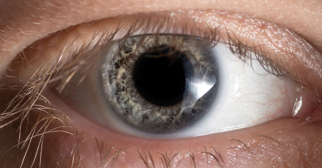
Whether you consciously notice it, or not, your eyes are constantly moving. Your eyes move quickly and reflexively from one point of focus to focusing on something else. During saccades—even if you aren't actively thinking about moving your eyes—the cerebellum is constantly taking inventory to pinpoint where to direct your focus, based on what holds the most important information. Finely-tuned saccades allow us to do things such as maintain eye contact with someone while engaged in conversation or volley a tennis ball zooming towards you at over 100 miles per hour.
Interestingly, one of the key symptoms of autism is having difficulty maintaining eye contact during emotional conversations. Autopsies of autistic children reveal shrunken cerebellums and atrophied Purkinje cells. I wrote about this phenomenon in a Psychology Today blog post, "What Inhibits Eye Contact During Emotional Conversations?"
The saccade is a perfect example of the cerebellum's sensorimotor coordination. How we coordinate all of our muscle movements has far-reaching impacts on how we interact with the environment and people around us. This is especially true when achieving a state of flow or superfluidity, which is marked by coordinating movements in a way that flows without any friction or viscosity.
The latest findings from OIST re-affirm my original hypothesis that Purkinje neurons play a pivotal role in achieving a state of flow and superfluidity both on-and-off the court. As I state on p. 119 of The Athlete's Way,
"You could think of one thousand purkinje cell dendrites as a “receiving dish” from many wide and varied places in your body. The single purkinje cell axon could be seen as one outgoing wire sending signals from all over the place through a consolidated pipeline. The dendrites of Purkinje cells are parallel but never touch. They oscillate like fishtails and push signals up the axon, out of the cerebellum, and up into the cerebrum."
This lightning-fast processing in a goalie’s Purkinje neurons in the cerebellum allows him to leap for a soccer ball while reaching out his hands and keeping his eyes locked on the target using the vestibulo-ocular reflex (VOR) of the cerebellum. In sport, and life, the cerebellum monitors your balance, proprioception, postural control, muscle coordination, and velocity of movements.

The final output of any given Purkinje cell is via a single axon. Interestingly, all the Purkinje cells appear to be working autonomously, but simultaneously in unison. These neurons take sensory information on a cerebellar level from every part of your body and send this information up to the cerebrum for cerebral processing.
The Purkinje neurons work at a quantum speed. The amplification of more than two hundred thousand incoming signals through one axon offers parallel processing capability from the cerebellum up into the cerebral cortex.
When the OIST team built a mathematical model on the average firing rate of the Purkinje neuron spikes, they found that a simple relationship could predict the fluidity of motion at the very beginning of a saccade eye movment.
Like all neurons, Purkinje cells emit spikes caused by electrical output. Purkinje cells fire spikes rapidly most of time, but occasionally there are pauses in these spikes. In a statement, Professor Erik De Schutter, co-author and head of OIST's Computational Neuroscience Unit, said.
"We want to know what these spikes are actually telling us . . . There is a big change in the local field potential at the time of a saccade. We can also see that there is a pause-beginning spike in the Purkinje cell at the time that the eye movement starts.
This showed us that the spikes that begin the pauses control the start of a movement and that the ones that are not related to the pauses control the velocity of the movement. This means that there is multiplexing in the Purkinje cells—they can send out two signals at once."
This is important because this research suggests that both timings of individual spikes and the average firing rate of the spikes are crucial to understanding the complexities of the cerebellum and all types of finely-tuned motor skills.
Conclusion: Achieving Superfluidity in Sport and Life Relies on Purkinje Neurons

As you watch elite-level athletes compete in the Rio Olympic summer games over the next few weeks, remember that all of their finely-tuned muscle movements rely on Purkinje neurons in the cerebellum. On the opposite end of the spectrum, atypical Purkinje cell structure, function, and connectivity can make it difficult for those with certain neurological disorders of the cerebellum to navigate everyday life.
These findings on Purkinje neurons have the ability to help experts develop interventions that could enhance an athlete's or artist's performance by creating flow and superfluidity. This research could also help those with a broad range of disorders that are rooted in cerebellar abnormalities optimize their daily lives.
Potentially, the latest insights into understanding the mechanism of the neurons in the cerebellum could lead to advances in medical technology—such as brain machine interfacing—which could allow paralyzed patients or amputees move prosthetics by fine-tuning brain signals. Additionally, these findings could be useful in designing advanced artificial intelligence for robots that require fine motor control.
Stay tuned for updates on ways cutting-edge cerebellar research can be applied to both sport and life. To read more on Purkinje cells, eye movements, and the cerebellum, check out my previous Psychology Today blog posts,
- "Purkinje Cells Burst to Life with State-Dependent Excitation"
- "Epigenetic Mechanism in the Cerebellum Drives Motor Learning"
- "The Whites of Your Eyes Convey Subconscious Truths"
- "The Neuroscience of Making Eye Contact"
- "12 Ways Eye Movements Give Away Your Secrets"
- "How Are Purkinje Cells in the Cerebellum Linked to Autism?"
- "Autism, Purkinje Cells, and the Cerebellum Are Intertwined"
- "More Research Links Autism and the Cerebellum"
- "Idiosyncratic Brain Synchronization Associated with Autism"
- "Enhanced Cerebellum Connectivity Boosts Creative Capacity"
- "The Cerebellum Holds Many Clues for Creating Humanoid Robots"
- "5 Reasons the Cerebellum Is Key to Thriving in a Digital Age"
© 2016 Christopher Bergland. All rights reserved.
Follow me on Twitter @ckbergland for updates on The Athlete’s Way blog posts.
The Athlete’s Way ® is a registered trademark of Christopher Bergland.


