Left Brain - Right Brain
Left Brain-Right Brain Research Isn't What It Used to Be
New studies shed light on how "left brain" and "right brain" rely on each other.
Posted February 9, 2021 Reviewed by Abigail Fagan

Two recently published studies advance our understanding of how "left brain-right brain" (i.e., the left and right cerebral hemispheres) work together to encode visual memories and how disproportionate deterioration of gray matter thickness in one cerebral hemisphere (but not the other) is associated with age-related cognitive decline and Alzheimer's disease.
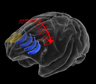
The first "left brain-right brain" study (Brincat et al., 2021) by researchers at MIT's Picower Institute for Learning and Memory investigated how the interhemispheric transfer of visual memories between the prefrontal cortex on the left and right side of the brain involves a coordinated process that allows mental images to "bounce between" the prefrontal cortex (PFC) in both cerebral hemispheres. This open-access paper, "Interhemispheric Transfer of Working Memories," was published on February 8 in the peer-reviewed journal Neuron.

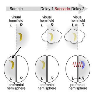
For this study, a team of Miller Lab researchers led by first author Scott Brincat and senior author Earl Miller, measured changing neural activity bilaterally using 256 electrodes placed on specific lateral regions ("hemisfields") of the left and right prefrontal cortex (PFC) as monkeys played a video game involving an image (e.g., banana or apple) that appeared and disappeared from the left and right side of a screen.
Miller's Lab also measured overall brain wave activity during the transfer of working memories between the left and right cerebral hemispheres and found that interhemispheric memory transfer "activates novel neural ensembles."
"As expected, [computer decoder] analysis showed that the brain encoded information about each image in the hemisphere opposite of where it was in the field of view," the authors explain in a news release. "But more remarkably, it also showed that in cases where the animals shifted their gaze across the screen, neural activity encoding the memory information shifted from one brain hemisphere to the other."
Notably, Brincat et al. "found that the transfer of a memory from one hemisphere to the other consistently occurred with a signature change in those rhythms." As the memory transfer occurred, synchrony of mid-range alpha/beta (~11-17 Hz) brain waves declined. On the flip side, during these memory transfers, synchrony of very low-frequency theta waves (~4-10 Hz) and high-frequency beta waves (~17-40 Hz) increased across both hemispheres.

"Around the time of transfer, synchrony between the two prefrontal hemispheres peaked in theta and beta frequencies, with a directionality consistent with memory trace transfer," the authors explain. "This illustrates how dynamics between the two cortical hemispheres can stitch together working memory traces across visual hemifields."
"Fast, reliable interhemispheric communication is critical for many real-world behaviors, including sports, driving, and air traffic control. Interhemispheric communication is also thought to be disrupted in some disorders, such as dyslexia," the authors conclude. "We hope that an understanding of the neural mechanisms of interhemispheric communication may lead to new ways to repair and optimize it."
Losing Gray Matter Volume in One Cerebral Hemisphere (But Not the Other) Has Surprising Ramifications
The second recent "left brain-right brain" study (Roe et al., 2021) was conducted by a team of Norwegian researchers from the University of Oslo who are part of the EU's Lifebrain project. Their open-access paper, "Asymmetric Thinning of the Cerebral Cortex Across the Adult Lifespan Is Accelerated in Alzheimer's Disease," was published on February 1 in Nature Communications.
Lifebrain is a European consortium that gives researchers in different countries shared access to comprehensive neuroimaging brain data from numerous age-related (0 to 100 years of age) longitudinal studies.
"The data we have thanks to Lifebrain is a treasure-trove. We were able to measure the thickness of every region of the cortex in over 2,600 healthy participants from five countries, up to six times in the same person over time," first author James Roe of the University of Oslo's Centre for Lifespan Changes in Brain and Cognition (LCBC) said in a news release. "Many other brain datasets only have one brain scan per person, so they cannot see changes occurring in the same person throughout life. Having follow-up scans of the same people was absolutely key to our study."
What did these comprehensive datasets of gray matter thickness reveal about changes in the left and right brain's cerebral cortex as people age? First, Roe and colleagues found that asymmetrical gray matter thickness ("cortical asymmetry") on the left and right side of the cerebral cortex was typical among healthy young people and seemed to improve cognitive functions.
"The left and right side of the cortex are not equally thick in younger brains," the authors explain. "Asymmetry is seemingly a good thing as it allows the brain to function optimally, as the left and right brain are specialized to do slightly different jobs."
As we get older, everyone loses some gray matter volume as the brain begins to atrophy and gradually "shrinks." However, Roe et al. found that, contrary to popular belief, the gray matter "bark" that encases the left and right cerebral hemispheres doesn't thin at the same rate on both sides of the brain during normal aging. (See, "4 Daily Habits That Could Stop Your Brain From Shrinking.")
Lifebrain's longitudinal datasets suggest that whichever side of the brain was thicker at age 20 deteriorates faster as people age; this negates the benefits of cortical asymmetry. "The asymmetry loss emerged at a similar age in most people (around their early 30s) and continued across the adult lifespan, with accelerated decline around age 60," the authors explain. "Loss of cortical asymmetry is happening gradually over the lifespan. We saw this with remarkable consistency in all samples," Roe added.
Lastly, James Roe and his University of Oslo colleagues found that the left side of the brain tends to shrink faster in Alzheimer's disease patients. "Overall, the present study may have unveiled the structural basis of a widely suggested system-wide decline in hemispheric specialization across the adult lifespan in brain systems subserving higher-order cognition, and found a potential continuation and acceleration of this decline in Alzheimer's disease," Roe et al. conclude.
Image credited to Meredith Mahnke for Miller Lab/MIT Picower Institute via EurekAlert
Graphical Abstract of Interhemispheric Transfer of Working Memories by Brincat et al., 2021/Neuron (CC BY-NC-ND 4.0)
References
Scott L. Brincat, Jacob A. Donoghue, Meredith K. Mahnke, Simon Kornblith, Mikael Lundqvist, Earl K. Miller. "Interhemispheric Transfer of Working Memories." Neuron (First published: February 08, 2021) DOI: 10.1016/j.neuron.2021.01.016
James M. Roe, Didac Vidal-Piñeiro, Øystein Sørensen, Andreas M. Brandmaier, Sandra Düzel, Hector A. Gonzalez, Rogier A. Kievit, Ethan Knights, Simone Kühn, Ulman Lindenberger, Athanasia M. Mowinckel, Lars Nyberg, Denise C. Park, Sara Pudas, Melissa M. Rundle, Kristine B. Walhovd, Anders M. Fjell, René Westerhausen & The Australian Imaging Biomarkers and Lifestyle Flagship Study of Ageing. "Asymmetric Thinning of the Cerebral Cortex across the Adult Lifespan Is Accelerated in Alzheimer's Disease." Nature Communications (First published: February 01, 2021) DOI: 10.1038/s41467-021-21057-y


