Autism
The Cerebellum, Cerebral Cortex, and Autism Are Intertwined
Neuroscientists have linked abnormal connectivity of the cerebellum with autism.
Posted April 10, 2015

Neuroscientists have identified a new marker for autism based on abnormal connectivity between the cerebellum and the cerebral cortex. This study is the first ever to look at connections between the entire cerebral cortex and the cerebellum using fMRI brain imaging.
Cognitive neuroscientists at San Diego State University (SDSU) recently discovered that in children and adolescents with autism spectrum disorder (ASD) the connections between the cerebellum and the cerebral cortex were overdeveloped in the sensorimotor regions of their brains.
On the flip side, study participants with autism had underdeveloped connectivity between regions of the brain involved in higher-order cognitive functions such as decision-making, attention, and language.
The April 2015 study, “Cerebro-Cerebellar Resting State Functional Connectivity in Children and Adolescents With Autism Spectrum Disorder,” was published in the journal Biological Psychiatry.
The study's first author Amanda Khan is a former master's student at SDSU and is currently a doctoral candidate at Suffolk University in Boston. Ralph-Axel Müller, a SDSU psychologist, is the study's corresponding author.
For this study, the researchers asked 56 children and adolescents—28 with autism and 28 without the disorder—to fixate on a focal point while thinking about nothing in particular while using fMRI brain imaging technology to scan the children's brains. Capturing this spontaneous activity allowed the researchers to hone in on the essential baseline neuronal patterns of each participant.

Typically, the sensorimotor connections between the cerebral cortex and cerebellum develop during the first few years of life. In a press release Müller said,
Our findings suggest that the early developing sensorimotor connections are highly represented in the cerebellum at the expense of higher cognitive functions in children with autism.
By the time the higher cognitive functions begin to come online, many of the connections are already specialized. If a particular part of the brain is already functionally active in one domain, there may be no reason for the brain to switch it over to another domain later in life.
During early childhood, scaffolding is laid down between various brain regions that creates the foundation for future neural networks that will either be pruned or strengthened through neuroplasticity. The key to learning and memory is based on the “fire and wire” principle that creates brain connectivity between brain regions that work together most efficiently.
The brain wants to be streamlined. Through plasticity, your brain cuts the connections between seemingly unnecessary networks and strengthens the connections between brain regions that require more robust lines of communication.

You can imagine the billions of neural connections within, and between, the cerebellum and the cerebral cortex like the old phone wires of a utility grid similar to these seen in this illustration of New York City from 1890.
It appears that in autism the overdevelopment of connectivity between the sensorimotor regions of the brain monopolizes neural networks and inhibits the connective wiring that is typically designated to serve higher cognitive functioning.
It's as if all of the communication lines in children with ASD are gobbled up by sensorimotor connections before the higher-order cognitive function connections have a chance to become a part of the brain’s other communication networks.
"Whatever the Cerebellum Is Doing, It’s Doing a Lot of It”
Traditionally, most neuroscientists have considered the cerebellum (Latin for “Little Brain”) to have the relatively simple job of overseeing muscle coordination and balance. Conventionally, neuroscientists don't give the cerebellum much credit for higher executive functions, cognition, psychiatric disorders, or emotional regulation. Luckily, these outdated notions of the cerebellum are rapidly evolving.
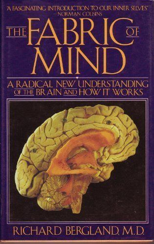
I have been dedicated to unraveling the mysteries of the cerebellum for over a decade. My father, Richard Bergland, was a neuroscientist, neurosurgeon, and author of The Fabric of Mind (Viking). He was obsessed with the cerebellum and passed this obsession on to me.

The cerebellum is only 10 percent of brain volume but holds over 50 percent of the brain’s total neurons.
Based on this disproportion, my father would always say, “We don’t know exactly what the cerebellum is doing, but whatever it’s doing, it’s doing a lot of it.”
My dad had a hunch that the cerebellum might play a role in cognitive function but was unable to prove this in his laboratory. He became frustrated by the limitations of 20th century brain imaging technology but, tragically, didn’t live to see the advent of the technology being used today.
When my father passed away in 2007, I made a vow that I would keep my antennae up for any new research about the cerebellum and do my best to vindicate his beliefs about the cerebellum posthumously in memoriam to him.
I have "Google Alerts" on my smartphone set to ding with a special sound whenever an update about the “cerebellum” or “cerebellar” is posted on the internet. I'm like a Pavlovian dog who salivates when I hear my phone ding with a Google alert that there's a news update about the cerebellum.
Needless to say, I was over the moon yesterday when I got the alert about this new study and was drooling all over the place. Again, this groundbreaking SDSU is the first ever to systematically look at connections between the entire cerebral cortex and the cerebellum using fMRI brain imaging.
Decreased Neural Activity Linked to Faster Learning
Interestingly, a different April 2015 study found that the fastest learners in a sensorimotor experiment actually showed decreased neural activity between specific brain regions in the frontal cortex and the anterior cingulate cortex.
This study was a collaboration that included University of California at Santa Barbara's Scott Grafton, M.D. and colleagues at the University of Pennsylvania and Johns Hopkins University. Their study, “Learning-Induced Autonomy of Sensorimotor Systems,” was published in the journal Nature Neuroscience.
In a press release Grafton described the research saying,
It's useful to think of your brain as housing a very large toolkit. When you start to learn a challenging new skill, such as playing a musical instrument, your brain uses many different tools in a desperate attempt to produce anything remotely close to music.
With time and practice, fewer tools are needed and core motor areas are able to support most of the behavior. What our laboratory study shows is that beyond a certain amount of practice, some of these cognitive tools might actually be getting in the way of further learning.
Grafton and his colleagues discovered that the visual and the motor blocks had a lot of connectivity during the first few trials, but as the experiment progressed they became essentially autonomous. Grafton explained, "the part of the brain that controls finger movement and the part that processes visual stimulus didn't really interact at all by the end of the experiment."
"Previous brain imaging research has mostly looked at skill learning over—at most—a few days of practice, which is silly," said Grafton. Adding, "Who ever learned to play the violin in an afternoon? By studying the effects of dedicated practice over many weeks, we gain insight into never before observed changes in the brain. These reveal fundamental insights into skill learning that are akin to the kinds of learning we must achieve in the real world."
Last month, I wrote a Psychology Today blog post titled, “The Cerebellum Deeply Influences Our Thoughts and Emotions,” based on the research that Jeremy D. Schmahmann, M.D is doing at Harvard Medical School.
Schmahmann has a theory that he calls “Dysmetria of Thought” which is basically a hypothesis that the cerebellum fine-tunes and coordinates our learning and thinking just like it fine-tunes and coordinates muscle movements.
With the idea of “dysmetria of thought” fresh in my mind, I emailed Dr. Scott Grafton to ask him if he thought that the cerebellum might be playing some role in why the quickest learners showed less neural activity. He responded, “Christopher, I don’t think the cerebellum could explain the complex changes of network communities we are observing. It is along for the ride, but there is no clear evidence it is driving these changes.”
As I try to connect the dots between all of the latest cerebellar research, I wonder if the findings from SDSU might dovetail in some way with Grafton’s research and help explain how the cerebellum is “along for the ride”?
The cerebellum remains very mysterious. I am optimistic that all of this research and discovery is part of solving a puzzle that might some day allow researchers to create revolutionary treatments for disorders like autism.
Conclusion: Superfluidity and the Positive Psychology of the Cerebellum
As with most neurological disorders, anything that takes someone “south of zero” on the -5 to +5 scale conversely has the ability to take someone “north of zero” when, instead of being dysfunctional, brain connectivity and plasticity are optimized.
I have a hypothesis that maximizing brain function and human potential can be achieved by optimizing the interconnectivity between each of the brain’s four hemispheres and finding ways to mitigate the detrimental plasticity of overconnectivity and underconnectivity.
Below is a rudimentary sketch I made a few years ago which illustrates my theory of "superfluidity" which is a term I use to describe the synchronicity of connectivity between the gray and white matter of every brain region within both hemispheres of the cerebrum and the cerebellum.

As an educated guess, I suspect that optimal brain function is obtained when all four brain hemispheres are working together in perfect harmony at an electrical, chemical, and architectural level.
I believe that a peak state of consciousness occurs when every nook and cranny of each of your brain's four hemispheres are working together in synchronicity. I call this a state of “superfluidity” because it represents absolutely zero friction, zero entropy and zero viscosity between thought, action, and emotion.
© Christopher Bergland 2015. All rights reserved.
The Athlete’s Way ® is a registered trademark of Christopher Bergland.


