Autism
Gray & White Matter MRI Opposite in Autistics vs. Psychotics
Imaging comparing autistic with psychotic brains confirms the diametric model.
Posted December 6, 2016
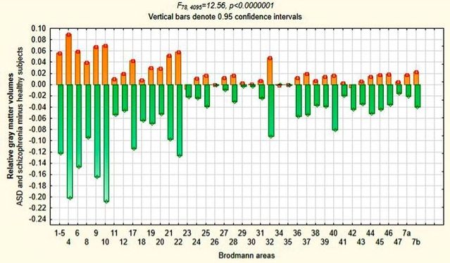
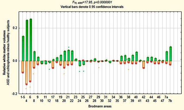
As I noted in a recent post, brain-imaging has finally begun to reveal fundamental differences in the brains of autistic as compared with psychotic subjects which contradict conventional wisdom but strikingly confirm the diametric model of mental illness. Now another study finds (to quote its title) a “Diametrical relationship between gray and white matter volumes in autism spectrum disorder and schizophrenia” in 49 subjects with schizophrenia, 20 subjects with autism and 39 healthy controls. Gray matter in the brain consists mainly of neurons, white matter mainly of neural connections. In the figures below, orange bars represent gray (above) and white matter volumes (below) in subjects with autism subtracted from volumes in healthy controls, and green bars the same for subjects with schizophrenia.


As the authors report, this study, aiming to compare subjects with schizophrenia and autism spectrum disorder (ASD),
unveiled a nearly identical topography but divergent vectors of volumetric changes in each group. We found a consistent pattern of volumetric differences between them in opposite directions in relation to healthy controls. Inverse relationships among changes in gray and white matter volumes were maintained for both patient groups: gray matter volumes were greater-than-normal in subjects with autism and lower-than-normal in subjects with schizophrenia; white matter volumes were greater-than than normal in subjects with schizophrenia and lower-than normal in subjects with autism. In relation to healthy controls, differences in volumes were more pronounced (and at least in white matter they were also more widespread) in subjects with schizophrenia than in subjects with autism. … This combination of overlap in topographical distribution of brain changes in subjects with ASD and schizophrenia with diametrically opposed volumetry of both gray and white matter could be seen as supportive of Crespi and Badcock (2008) theory proposing that ASD and schizophrenia represent opposite extremes of the same phenotypic spectrum. As a corollary, our findings are inconsistent with the view that relationship between ASD and schizophrenia is predicated on mere overlap in their impairment of social brain.

The authors go on to add that they “found no areas of increased gray matter volumes in subjects with schizophrenia and decreased gray matter volumes in subjects with autism, and consequently uniformly greater gray matter volumes in autism than schizophrenia,” which they think is “difficult to reconcile with the Crespi and Badcock hypothesis.” But in The Imprinted Brain, I commented that "autism and schizophrenia exhibit divergent patterns of grey matter loss, with little or no loss in autism, moderate loss in normal development, and high rates of loss in schizophrenia" (p. 166), just as illustrated top left. I also cited the suggestion that global under-connectivity goes with local hyper-connectivity in autism; with the diametrically opposite situation—local under-connectivity with global hyper-connectivity—being the case in schizophrenia: something which obviously fits the diametric model (p. 104). If this translates into more white matter than normal in schizophrenics (because global hyper-connectivity necessitates much longer connections than does local hyper-connectivity), the result would nicely fit the finding illustrated bottom left above. And as the authors themselves point out, "localized regional increases in gray matter volumes may be sufficient to explain the prevalence of autistic savant abilities in isolated cognitive domains." Furthermore, I would argue that enhanced global connectivity correspondingly seems to explain much about psychotic hyper-mentalism: notably paranoid, magical, and grandiose/religious delusions.
But whatever else you might say, the pattern revealed in Figure 3 as a whole is one of mirror-image symmetry of autism and schizophrenia: exactly what you would expect if you took the diametric model to be applicable to both white and gray matter. As the authors themselves remark, "In conclusion, this study supports diametrical positioning of autism and schizophrenia along the same dimensional spectrum." And given that, as they also point out, "no prior study attempted a systematic assessment and parcellation of white matter volumes throughout the brain," their findings add up to one of the most remarkable vindications so far of the diametric model—not to mention refutations of conventional wisdom.
Finally, there are important implications for diagnosis as the authors imply in their report that they "did not find statistically significant differences in gray or white matter volumes between subjects with autistic and Asperger’s disorders, thus supporting the elimination of this nosological distinction in DSM-5." Up to now, MRI (magnetic resonance imaging) has been widely used in medicine for definitive diagnosis of disorders like tumors in the brain, but has been of limited use in psychiatry otherwise. The reason is that conventional psychiatry does not know what to look for or where to look for it. But if these findings are confirmed, they imply that MRI could now become an important tool for psychiatric diagnosis, notably for resolving one of the thorniest issues which besets the diametric model: that of the apparent overlap of autistic and psychotic symptoms which was the subject of some recent posts on this site. Clearly, if autistic, as opposed to psychotic spectrum disorders could be distinguished by MRI as this landmark study suggests they might, psychiatric diagnosis would be placed on a sound factual basis, visibly revealed in the brain.
(With thanks to Amar Annus for bringing this to my attention.)


