Cognition
Alzheimer's Study Links Triad of Brain Areas with Cognition
Atrophy in cortical, subcortical, or temporal brain areas linked to Alzheimer's.
Posted October 7, 2016

A new study published online today in Proceedings of the National Academy of Sciences reports that distinct patterns of atrophy within three different brain areas (cortical, subcortical, or temporal) might help to explain how the loss of various cognitive abilities manifests in patients diagnosed with Alzheimer's disease (AD).
For this study, a team of researchers from Massachusetts General Hospital (MGH) at Harvard Medical School and the National University of Singapore (NUS) used mathematical modeling to pinpoint how various brain regions—some of which are not typically linked to cognition—may impact cognitive abilities.

As an example, the cerebellum (Latin for "little brain") has historically been thought of by the medical establishment as being responsible solely for 'non-thinking' activities such as fine-tuning muscle coordination. This new research suggests that the cerebellum may, in fact, play an important role in cognitive function or the degeneration of executive function and memory.
In a statement to Massachusetts General Hospital, Thomas Yeo, Assistant Professor at the NUS Department of Electrical and Computer Engineering and the MGH Laboratory for Computational Neuroimaging, said,
"The symptom severity and neurodegeneration can vary widely across patients in Alzheimer's disease. Our work shows that participants in this study exhibit at least three atrophy patterns—cortical, temporal or subcortical—that are associated with variability in cognitive decline not only in patients diagnosed with Alzheimer's but also in individuals with mild cognitive impairment or those who are cognitively normal but are at risk for Alzheimer's."
Yeo’s research focuses on the development of machine learning algorithms for the large-scale analysis of high-dimensional and complex brain imaging data. These mathematical computational models allow researchers to characterize how various brain systems support cognition.
For this recent study, Yeo and colleagues analyzed data that was gathered as part of the Alzheimer's Disease Neuroimaging Initiative (ADNI). The study included a total of 379 participants: 188 had been diagnosed with Alzheimer's disease, 147 with mild cognitive impairment, and 43 who were not cognitively impaired but at a higher risk for beta-amyloid plaques that are associated with developing Alzheimer's disease.
As a first step to creating mathematical models, the research team analyzed data from baseline structural MRIs. These neuroimages helped to estimate the probability that atrophy in a particular brain region was linked to specific changes in executive function and memory. Based on the location of atrophy factors, the researchers determined a correlation associated with a combination of three different atrophy patterns.
Three Atrophy Patterns and Brain Areas Associated with Alzheimer's Disease
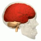
- Cortical: Indicating atrophy throughout the cerebral cortex (Brain areas in the cerebrum, Latin for "brain").
- Subcortical: Indicating atrophy in the cerebellum, striatum, and thalamus. (Brain areas at the base of the brain).
- Temporal: Indicating atrophy of the medial temporal cortex, hippocampus, and amygdala (Brain areas in the cortical lobe behind the ears).
All three brain areas were associated with executive function and memory decline across the entire clinical spectrum. The cortical factor was most strongly associated with executive function decline. The temporal factor showed the strongest association with memory. The subcortical factor was associated with the slowest decline of both executive function and memory. These findings suggest that distinct patterns of brain atrophy can be linked to various cognitive domains.

As is always the case, analyzing the brain mechanics of any neurological disease or disorder helps to pinpoint how changes in brain structure or functional connectivity are linked to specific cognitive abilities.
For example, in 1848, Phineas Gage suffered an accident in which an iron rod pierced through his orbitofrontal cortex (OFC) in the frontal lobes. Before his brain injuries, Gage was known to be a congenial and soft-spoken man who lived by the rules of society. After his accident, Gage became an uninhibited, and often temperamental nonconformist who paid little regard to the rules of society and could be adversarial. He lived until 1860, but after the iron-rod incident, his personality was so altered that his friends and family referred to him as "no longer Gage." The direct link between OFC damage and personality changes in Gage helped researchers identify the function of this particular brain region.
Just as neuroscientists pinpointed the role of various lobes in the cerebrum during the 20th century, the next frontier in neuroscience during the 21st century will be to map how various "micro-zones" throughout the entire brain interact with one another. The discovery of how various combinations of brain atrophy in three brain areas is linked to Alzheimer's helps to advance our understanding of how changes in gray matter volume in different brain regions affects specific aspects of cognition.
"Dysmetria of Thought" Hypothesizes that Subcortical Regions Play a Role in Cognition
Along these lines, Jeremy Schmahmann (also of Massachusetts General Hospital) believes that the posterior cerebellum could take center stage in the future based on its significant role in our human evolution and findings from his MGH ataxia unit. The posterior lobe has expanded exponentially in our recent evolution. In fact, only the prefrontal cortex has grown more rapidly than the posterior cerebellum during our evolution.
Schmahmann has a hypothesis he calls "Dysmetria of Thought" which is a theory that the cerebellum may fine-tune our thoughts just as it fine-tunes our muscle movements. He developed this theory after studying multiple patients with damage to the cerebellum and observing a pattern of deficits in the cognitive domains of executive function, spatial cognition, and language. The new research by his colleagues in the computational neuroimaging lab at MGH, affirms that subcortical regions (including the cerebellum) play a role in executive function and memory.
In a 2010 study, Schmahmann and his colleagues at MGH coined the term Cerebellar Cognitive Affective Syndrome (CCAS) which is also referred to as "Schmahmann's Syndrome.” CCAS is represented by impairments of executive function that include problems with planning, set-shifting, abstract reasoning, verbal fluency, and working memory. The symptoms of CCAS are perseveration, distractibility, and inattention which are all correlated with lesions, damage, or atrophy of the cerebellum.
Interestingly, the analysis of brain scans in the recent study by Yeo et al. indicated that various atrophy factor patterns two years after the initial scans were persistent in a wide range of individuals. Most participants—including those in the mild cognitive impairment and cognitively normal—showed levels of more than one brain area that had atrophied.
"Most previous studies focused on patients already diagnosed, but we were able to establish distinct atrophy patterns not only in diagnosed patients but also in at-risk participants who had mild impairment or were cognitively normal at the outset of the study," Yeo says. "That is important because the neurodegenerative cascade that leads to Alzheimer's starts years, possibly decades, before diagnosis. So understanding different atrophy patterns among at-risk individuals is quite valuable."
Future Research Will Explore How Various Brain Regions Impact Other Neurological Disorders
More research is needed to better understand how specific atrophy patterns are related to the distribution of amyloid and tau and to pinpoint the exact mechanism through which these changes impact specific cognitive abilities. In his statement to MGH, Yeo concluded,
"Previous studies assumed that an individual can only express a single neurodegenerative pattern, which is highly restrictive since in any aged person there could be multiple pathological factors going on at the same time—such as vascular impairment along with the amyloid plaques and tau tangles that are directly associated with Alzheimer's. So individuals who are affected by multiple, co-existing pathologies might be expected to exhibit multiple atrophy patterns."
Thomas Yeo emphasizes that the same type of mathematical modeling used to analyze neuroimaging data in this study on Alzheimer's can be used to deconstruct the role that various brain regions play in other neurological disorders. Future research will explore how a variety of clinical symptoms and gray matter volume patterns play out in other brain disorders such as Parkinson's disease, schizophrenia, and autism. Stay tuned!


