Autism
Autism, Purkinje Cells, and the Cerebellum Are Intertwined
Abnormal Purkinje cell neuroplasticity is linked to autism spectrum disorder.
Posted November 25, 2014

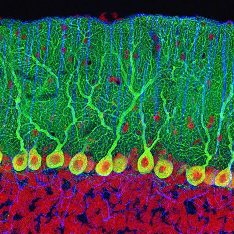
Purkinje cells of the cerebellum.
A new study from the University of Chicago Medical Center has identified that malfunctioning Purkinje cell neuroplasticity is directly associated with the reduced capacity for motor learning in children with autism spectrum disorder (ASD).
The November 2014 study, “Cerebellar Plasticity and Motor Learning Deficits in a Copy-Number Variation Mouse Model of Autism,” is published in the journal Nature Communications.
Purkinje cells and the cerebellum are the prime driving force of The Athlete’s Way. I have had my antennae up for new research about the role of the cerebellum and Purkinje cells in life and sport for over a decade.
I was excited last week to see the cerebellum take center stage at the 44th annual Society of Neuroscience meeting in Washington, D.C. Many researchers at the conference put Purkinje cells in the spotlight.

Cerebellum in red.
In 1504, Leonardo da Vinci made wax castings of the brain and coined the term “cerebellum” which is Latin for “little brain.” The cerebellum is only 10% of brain volume but holds over 50% of your brain’s total neurons.
Purkinje cells are named after Johannes Purkinje, who first identified these neurons in 1837. Dr. Purkinje was also the first person to identify the individuality of the human fingerprint. Among many functions, the Purkinje cells—which are held in the cerebellum—are responsible for fine-tuned motor control, balance, proprioception, vestibulo-ocular reflex (VOR) and communicating information from the cerebellum to the cerebral cortex.

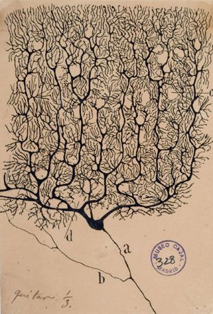
Purkinje cell drawing by Santiago Ramón y Cajal.
Almost 80 percent of children with autism spectrum disorder have problems with coordination, motor control, and focusing their eye movements. Until recently, the underlying cause of these motor deficits in children with ASD was an enigma.
In a fascinating study, the neuroscientists at the University of Chicago used ASD model mice to identify the abnormal connections between Purkinje cells which they believe are the likely cause of motor coordination issues seen in autism spectrum disorder.
In a press release, study senior author Christian Hansel, PhD and professor of neurobiology at the University of Chicago said, "We have identified synaptic abnormalities that may play a role in motor problems typically seen in children with autism. Autism is sometimes described as intense world syndrome—too many, too strong excitatory connections that lead to enhanced sensory input. The results of our study might shed light on this phenomenon."
Of Mice and Men: Mouse Models of ASD Offer Clues for Humans with Autism
Hansel points out, "A direct link between synaptic studies and behavioral output is almost impossible to do with social behaviors, but we can now accomplish this. This is due to the relative simplicity of the motor system, and because the cerebellum is evolutionarily conserved, allowing for comparisons between mice and man."
For this study, Hansel and his team used mice that have a common genetic abnormality (patDp/+) found in autism which is the human 15q11-13 chromosomal duplication.
The researchers found ASD model mice demonstrated motor deficits in the form of an unstable gait and impaired motor learning. The researchers used classical conditioning to train all of the mice to associate a short light signal with a puff of air to the eye which would cause them to blink. The researchers timed the response times within milliseconds.
All of the mice became conditioned to blink in response to the light, even when the air puff was removed. However, the researchers found that the ASD model mice were much slower to have the conditioned blinking response hardwired into muscle memory and made mistakes more often than the other mice.
To understand why this was happening, the researchers honed in on the Purkinje cells which are directly linked to muscle memory and motor learning.
Purkinje Cells Are Why Practice Makes Perfect

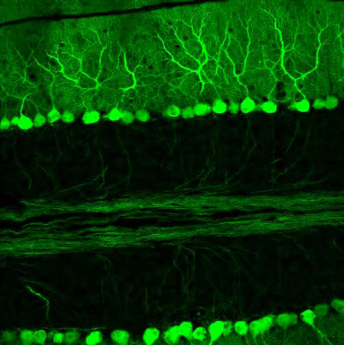
Purkinje cells in green.
Purkinje cells can either strengthen or depress the efficacy of their synapses. The inhibition of Purkinje cells comes through practice and conditioning and is the key to mastering complex fine-tuned motor skills used when playing a musical instrument, serving a tennis ball, performing surgery, etc.
On page 119 of The Athlete's Way I describe the unique structure of the Purkinje cells as I learned it from my father, Richard Bergland, who was a neuroscientist and neurosurgeon:
The syanptic plasticity of Purkinje cells is in your hands. They are reshaped daily through practice and repetition. The Purkinje cells work at a quantum speed. The amplification of more than two hundred thousand incoming signals through one axon offers parallel processing capability from the cerebellum to the cerebrum ... The final output of any given Purkinje cell is via a single axon but all the Purkinje cells are working autonomously, but simultaneously. They oscillate together and march in lock step. These cells take sensory information from all parts of the body and send it to the cerebrum.
In ASD model mice, the University of Chicago researchers found that the ability of Purkinje cells to depress the efficacy of their synapses was greatly reduced. The researchers concluded that this hindered their ability to help fine-tune the muscle movements needed for efficient motor learning.

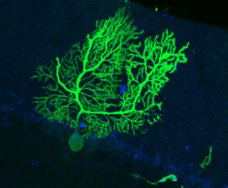
Purkinje cell in green.
They discovered that a likely cause of the hyperactivity of the Purkinje cells was linked to impaired synaptic pruning. The key to optimizing neural plasticity is always going to be the balance of neural darwinism and pruning unneeded synapses combined with the "fire-and-wire" of necessary synaptic connections. Ideally, synapses that fire together wire together and those that are not used regularly are pruned.
Purkinje cells rely on connecttions with neuronal projections called climbing fibers to receive an infinite number of environmental updates that keep the cerebellum and cerebrum aware of when things are going right, and when there are errors or disturbances.
Signals from climbing fibers can trigger an appropriate motor response in milliseconds. When pruning is working normally, each Purkinje cell in an adult brain receives only one climbing fiber input. ASD model mice, however, possessed an overabundance of climbing fibers which made it more difficult to interpret an 'error signal.'
"Inefficient synaptic pruning seems to be a common motif in autism," Hansel said. "There are not many types of synapses in the brain where pruning can be measured easily, but climbing fibers provide an excellent model and allow us to make predictions about synaptic pruning deficits elsewhere in the brain as well."
Conclusion: More Research Is Needed on Purkinje Cells and Climbing Fibers
Hansel and his team have demonstrated that synaptic abnormalities in the Purkinje cells are associated with the delayed eye blink response seen in ASD model mice. This is a breakthrough discovery.
Moving forward, autism researchers can use this understanding of the neural circuitry within the cerebellum to develop interventions and treatments for people with autism spectrum disorder.
The researchers hope that their findings will help pave the way for clinical trials that focus on the treatment of synaptic dysfunction of Purkinje cells and climbing fibers.
If you'd like to read more about Purkinje cells, the cerebellum, and the implicit learning of muscle memory check out my Psychology Today blog posts:
- "How Are Purkinje Cells in the Cerebellum Linked to Autism?"
- "Research Links Autism Severity With Motor Skill Deficiencies"
- "The Neuroscience of Knowing Without Knowing"
- "Is Cerebellum Size Linked to Human Intelligence?"
- "Primitive Brain Area Linked to Human Intelligence"
- "The Mysterious Neuroscience of Learning Automatic Skills"
- "Too Much Chrystalized Thinking Lowers Fluid Intelligence"
- "The Neurobiology of Grace Under Pressure"
- "How Does the Vagus Nerve Convey Gut Instincts to the Brain?"
- "Why Does Overthinking Cause Athletes to Choke?"
- "Toward a New Split-Brain Model: Up Brain-Down Brain"
- "Neuroscientists Discover How Practice Makes Perfect"
- "How Is the Cerebellum Linked to Autism Spectrum Disorders?"
- “Childhood Family Problems Can Stunt Brain Development”
- "The Neuroscience of Calming a Baby"
- “Why Is Dancing So Good For Your Brain?”
- “The Neuroscience of Madonna’s Enduring Success”
- "Gesturing Engages All Four Brain Hemispheres"
- "The Neuroscience of Superfluidity"
- "One More Reason to Unplug Your Television"
- "Better Motor Skills Linked to Higher Academic Scores"
- "Hand-Eye Coordination Improves Cognitive and Social Skills"
- "The Neuroscience of Imagination"
- "Can Practice Alone Create Mastery?"
- "No. 1 Reason Practice Makes Perfect"
Follow me on Twitter @ckbergland for updates on The Athlete’s Way blog posts.
Photo Credits:
- Thomas Deerinck/ Tumblr
- Wikimedia Commons


