Fear
Brain Scanning Enables an Objective Look at Animal Emotions
Research reveals how the brain of a crow assesses dangerous situations.
Posted September 15, 2012
Anyone who spends time in the close company of a pet knows how to read the emotions of another blood. When my border collie cowers, I know she is afraid, and when she bows, head down, butt up with tail wagging, I know she is playful. Reading the emotion in wild animals is more difficult, but this does not mean they are not emotional. In fact, a study just published by the Proceedings of the National Academy of Sciences (www.pnas.org) shows how we used brain scanning technology to document the emotions of fear and expectation in wild crows. By partnering with colleagues in the Department of Radiology at the University of Washington, we were able to visualize the activity in crows’ brains as they viewed a dangerous person (the person who originally captured them) and a caring person (the person who fed them in captivity). What we found was familiar; the crows used the same parts of their brains when confronting a known danger as we do. Because similar parts of the brain were activated, and similar brain chemicals (neurotransmitters and hormones) control this activation in birds and mammals, including humans, I think it is no stretch to also conclude that crows felt as we might when we see a dangerous person. They were scared and anxious. In fact, they froze, like a deer in a car’s headlights or a person staring at an armed bandit, when confronted by their former captor. While these results were very interesting and satisfying to me as a scientist, the technique my colleagues brought that enabled our discovery was even more important. The PET scanning approach we used, while requiring us to capture and briefly hold crows in captivity, was minimally invasive. After concluding our tests, the birds could be released back to the wild. Opening the door to the carrying cage to set the birds free again on their home turf was the best part of this research. As each left the cage they flew high and strong. And I left gratified for the chance to know each on a personal level, having seen their brains in action.
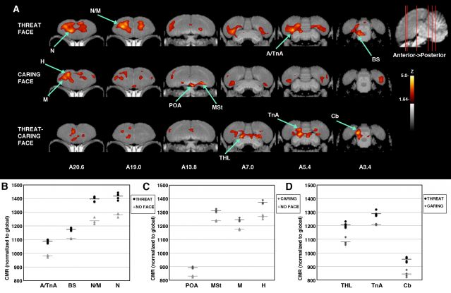
Tony and I wrote about this research in Gifts of the Crow. I paste in our text about the research below, and I include an image of crow brains from the recent scientific article that shows the different brain regions used by crows as they view a threatening (the person that captured them) and a caring (the person who fed them) person. The first two rows in the image show the areas in the crow brain that were activated by the sight of a threatening (top row) and caring (second row) person. The difference in these areas of activation is shown in the third row. The actual amount of activity is shown for each labeled region in the small graphs (regions are given letters on the images: N=nidopallium, M=mesopallium, A=arcopallium, TnA=Amygdala, MSt=striatum, BS=brainstem, THL=thalamus, H=hyperpallium).

Image of crow brain activity while viewing a dangerous and a caring person (top two rows). Most active areas are colored.
"While observing behavior tells us something about how crows view us, it tells us very little about how their brains reacted when they saw us. That all changed when John had the good fortune of meeting a few new colleagues at the University of Washington Medical Center. Dr. Donna Cross, Dr. Robert Miyaoka, Barbara Lewellen, and Greg Garwin specialize in the imaging of animal subjects. They routinely scan the bodies and brains of monkeys, rats, and mice to understand brain function, cancer, post traumatic stress, autism and other human afflictions. When Heather Cornell, a student who conducted our studies of human recognition, and John met Donna, Robert, Barbara, and Greg they had never peeked inside the brain of a bird. But they were game to try.
We decided to make the first attempt to measure how a crow’s brain reacted to the sight of a dangerous person. Heather and John caught thirteen crows wearing one of our custom-made, realistic masks. As our birds acclimated to their individual, outdoor flight cages, we fed and tended them while wearing a different mask. By keeping their contact with other people to a minimum over the next few weeks, we would be able to compare their reactions when they glimpsed a familiar-but-dangerous versus a familiar-but-caring face. We were ready to look at their brains in action. The procedure Donna and her team proposed is called PET (positron emission tomography) scanning, widely used in people and animals and in order to visualize where the brain was active in the half hour or so before the scan is done. This ability to look back at a crow’s mental activity is important because during the scan the bird must hold still, which requires anesthesia. We also used the more familiar scanning technique, MRI, but because this technique scans what is currently happening, not what has happened, we used it only to get a detailed look at the structure, not function, of the crow’s brain.
Here is how we scanned the crows’ brains: We brought a test crow up to Robert’s lab and allowed it to get used to a new, smaller cage for an evening. The next morning John reached through a curtain, grabbed the crow, blindfolded it, and gave it a small injection of radioactively labeled glucose. John then put the bird back, playing soothing crow calls for a few minutes, and Heather and he removed the curtain to allow the bird to see their faces. Some of the birds saw them wearing the mask worn when they were captured, some saw the mask worn when they fed them and cleaned their outdoor cages, and a few saw no person, only the room. After fifteen minutes of on and off viewing, Greg and Barbara anesthetized the bird, wrapped it in a warm blanket, monitored its vitals, and slid it into the PET scanner. As the crow slept, the scanner mapped the occurrence of the radioactive tracer within its brain. As the bird was viewing Heather and John, its whirring brain was demanding energy that was supplied in part by the labeled glucose they had slipped it. The most active parts of the brain would require the most glucose and therefore receive the greatest label; less would accumulate where the brain was less active. The scanner, like a 3D x-ray machine, would paint us a picture of the tracer’s deposition. In an hour we would know how the crow was thinking. In a day, after clearing its body of the tracer, we could free the crow and let it resume its life in the wild.
As the first crow awakened from his isoflurane-induced sleep, Robert was already busy reconstructing an image of the bird’s brain. Amazingly, we had a computer rendition of the crow’s brain that we could explore bit by bit. Right away we saw the eye’s retinas, the thalamus, and the entopallium blazing with activity—this was the trace of visual information streaming into the crow. There was also activity scattered throughout the forebrain. Our crow had taken in the scene. But would the brain reflect anything about whom the crow saw or would it just reflect fear of a strange place, a sudden grab, and a shot in the belly? Donna next worked on the images to compare, point by point, the brains of crows that had viewed dangerous, safe, and no faces.
The results were striking. As in human images, we saw a complex network of brain regions respond to our presence. Sensory areas in visual pathways translated sight into neural activity. The integrative nidopallium and mesopallium, and the associative striatum were active, as expected if our crows were evaluating their visual experience in the context of memory. When looking at a person, crows used one side of their brains more than the other, just as sheep do; their left hippocampus and right nidopallium were especially active. And some areas appeared especially tuned to the dangerous face. When viewing a dangerous face, our crows used their nidopallium, arcopallium, amygdala, and areas in their thalamus and brainstem known to be important to fear responses. This reaction was remarkably similar to that of a person who views a dangerous situation. Our crows even relied mostly on the right hemisphere of their brains, just like people do in fearful settings.
The activity in the brain of a crow who looked upon a caring person was quite different from that of a crow who saw a dangerous person. Upon seeing a caring face, the preoptic area and striatum of the brain were most active. These regions are known to be part of the social brain network stimulated during social interactions, where their activity indicates a bird’s hunger and its attention to learned associations. This suggests that crows perceived the association they learned between food and their human caretakers. Again, our crows even varied the use of their two brain hemispheres, exactly as do humans. Instead of using their right brain, as was the case when seeing danger, now they used their left brain. Clearly, as with humans, crows pay attention to peoples’ faces and integrate what they see with what they remember and feel, using a complex neural circuit to evaluate each of us."
(Gifts of the Crow, Free Press, Copyright 2012 by John Marzluff and Tony Angell)

