Autism
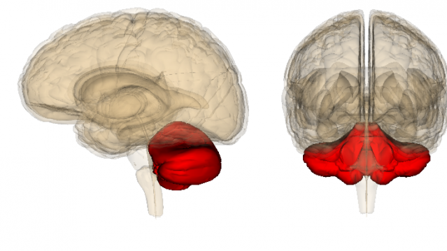
More Research Links Autism and the Cerebellum
Growing evidence links autism spectrum disorders and the cerebellum.
Posted August 16, 2015

A new study led by Samuel Wang, professor of molecular biology at Princeton University, suggests that abnormalities of the cerebellum are correlated with some of the sensory difficulties seen in autism spectrum disorders (ASD). Until now, sensory integration difficulties have been reported in autism, but their underlying brain-circuit mechanisms have remained enigmatic.
In previous Psychology Today blog posts, I've written extensively about the research that Sam Wang and colleagues have been conducting on the cerebellum. This entry is an update to a November 2014 post I wrote titled, "How Are Purkinje Cells in the Cerebellum Linked to Autism?"
The new study by Wang et al used five different autism-related mouse models—each with a different genetic mutation that has previously been linked with autism—to help the team of researchers identify specific correlations between each mutation and various types of delayed eyeblink conditioning. Mice carrying any one of five autism-linked mutations all have difficulty associating a flash of light and a puff of air during classical eyeblink conditioning.
The July 2015 study, "Cerebellar Associative Sensory Learning Defects in Five Mouse Autism Models,” was published in the journal Elife. Cerebellar is the sister word to "cerebral." Cerebellar means relating to or associated with the cerebellum.
How Is the Cerebellum Associated With Autism Spectrum Disorders?
Autism spectrum disorders in humans are typically characterized by social deficits, communication difficulties, repetitive behaviors, sensory information issues, and in some cases, cognitive delays.
Until now, it was puzzling which circuits in the brain are responsible for various differences in eyeblink conditioning and autism. The new study breaks new ground by asking whether specific versions of genes that increase the risk of autism in humans also disrupt eyeblink conditioning in mice.
The new findings suggest that ‘autism’ mice have trouble receiving and integrating sensory information from multiple senses, a brain function that is regulated by the cerebellum. In a press release, Wang said, “Many people with autism have difficulty integrating information from multiple senses. The air puff test may model this sensory struggle.”
The cerebellum receives multitudes of sensory information and translates this input into coordinated actions. Previous brain imaging and postmortem studies have found abnormalities in the cerebellum of people with autism. However, exactly how these abnormalities relate to autism symptoms remains unclear.
Researchers Use Eyeblink Conditioning to Link Autism and the Cerebellum
When a sensory perception test involving cerebellar reflexes is performed in a laboratory, the most widely used measurement is a form of reflexive learning or classical conditioning called “eyeblink conditioning.”
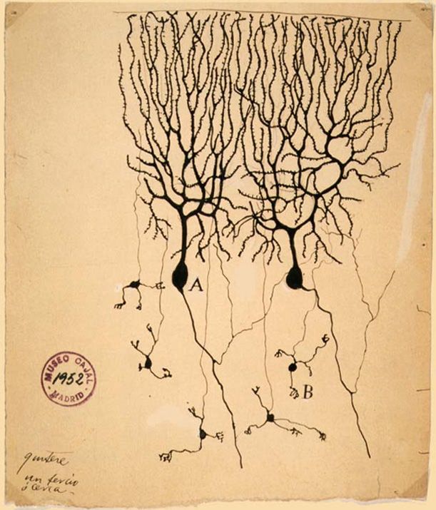
The eyeblink conditioning process is relatively simple. It consists of pairing an auditory or visual stimulus (the conditioned stimulus (CS)) with an eyeblink-eliciting unconditioned stimulus (US) such as a mild puff of air to the cornea. In both mice and humans, the eyblinking reflex relies on the cerebellum, and becomes hardwired in cerebellar neurons that include granule and Purkinje cells. In a press release, Wang stated:
Some people with autism also have trouble anticipating a puff of air. Performing this test in young children may reveal problems with sensory integration before other autism symptoms appear. If you think about it, all early-life learning is multisensory What else do babies do other than link the taste of milk with mom's voice?
For this experiment, the researchers affixed a tiny magnet to the lower eyelid of each mouse and also placed a detector on the upper eyelid to measure how quickly and fully each mouse blinked with the combination of a flash of light and a puff of air. Of the five different autism mice mutations, eyeblink differences fell into two distinct categories: 1) Eyeblinking problems with associating the flash of light with the puff of air. 2) Translating the light and puff of air into an eyeblinking action.
More specifically, the researchers state, “Mice missing TSC1 never learn to anticipate the puff of air, and the SHANK3, 15q duplication and CNTNAP2 mice anticipate it significantly less often than controls do. Mice with mutant MeCP2 learn to blink, but their timing is off: They close their eyes too late and less completely than controls. SHANK3 and TSC1 mutant mice also close their eyes less than controls, but SHANK3 mice appear to blink a little too early.”
Conclusion: Receiving and Integrating Sensory Information Relies on Purkinje Cells and Granule Cells in the Cerebellum

The latest study by Wang and colleagues shows that the probability of typical eyeblink conditioning was reduced by various mutations in the cerebellum. By identifying how specific mutations are linked to various types of abnormal learned responses during eyeblink conditioning, the researchers move one step closer to solving the riddle of exactly how the cerebellum is linked to autism.
In the press release, Matt Mosconi, assistant professor of psychiatry at the University of Texas, Southwestern in Dallas said, "The real importance of this study is that it suggests the severity and nature of the cerebellar alterations may vary across the different models.”
More specifically, the SHANK3 and MeCP2 are expressed in granule cells—neurons in the cerebellum that receive sensory signals. Therefore, these findings suggest that granule cells are part of a circuit that controls the timing of blinks. On the flip side, TSC1 is expressed in Purkinje cells, which belong to a circuit that helps integrate sensory information.
It appears that receiving and integrating sensory information relies on optimal function of both the granule cells and Purkinje cells. When there are abnormalities in these various cells, autism-related sensory problems may occur to varying degrees.
The authors conclude, “Overall, our observations are potentially accounted for by defects in instructed learning in the olivocerebellar loop and response representation in the granule cell pathway. Our findings indicate that defects in associative temporal binding of sensory events are widespread in autism mouse model.”
To read more about the cerebellum, check out my previous Psychology Today posts:
- "The Cerebellum Deeply Influences Our Thoughts and Emotions"
- "The Cerebellum, Cerebral Cortex, and Autism Are Intertwined"
- "Autism Genes Can Disrupt Connections Between Brain Regions"
- "How Are Purkinje Cells in the Cerebellum Linked to Autism?"
- "Autism, Purkinje Cells, and the Cerebellum Are Intertwined"
- "Research Links Autism Severity With Motor Skill Deficiencies"
- "How Does Your Cerebellum Counteract 'Paralysis by Analysis'?"
- "How Does Practice Hardwire Long-Term Muscle Memory?"
- "Neuroscientists Discover How Practice Makes Perfect"
- "Want to Improve Your Cognitive Abilities? Go Climb a Tree!"
- "The Cerebellum May Be the Seat of Creativity"
© 2015 Christopher Bergland. All rights reserved.
Follow me on Twitter @ckbergland for updates on The Athlete’s Way blog posts.
The Athlete’s Way ® is a registered trademark of Christopher Bergland.


