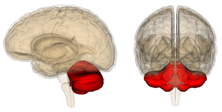
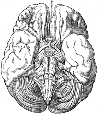
Left Brain - Right Brain
My Decades-Long Quest to Decode a Quirky “Super 8” Brain Map
Like an arcane treasure map, this hand-sketched brain map holds many mysteries.
Posted February 19, 2021 Reviewed by Kaja Perina

An overview of how the structure and connectivity of both cerebellar hemispheres affect motor and nonmotor functions of the cerebral cortices.
Since 2009, I've been on a mission to solve the riddles held in a brain map (shown in color halfway down the page) that I hastily scribbled down one afternoon using different colored Sharpie markers and Highlighters from an office supply store.
The origin story of my hand-sketched map begins in the early 1970s, when my late neuroscientist father, Richard Bergland (1932-2007), who wrote The Fabric of Mind, repeatedly told me during weekend tennis lessons that our so-called "muscle memory" is held in the cerebellum's Purkinje cells. "Think about hammering and forging the muscle memory into your cerebellum with every stroke," Dad would say.
Because the cerebellum is well known for coordinating muscle movements, it made sense that my father viewed optimizing cerebellar functions as vital to an athlete's peak performance. When I was writing The Athlete's Way in the mid-2000s, I consulted with my father daily; together, we created a split-brain model called "up brain-down brain." The main objective of this framework is to facilitate flow states by minimizing cerebral overthinking and fine-tuning automatic, non-thinking cerebellar movements. (See "The Split-Brain: An Ever-Changing Hypothesis.")

Although our split-brain model was straightforward and the simple "upstairs (cerebrum) vs. downstairs (cerebellum)" duality was easy to convey, it only told half the story. The real twist to how the cerebrum and cerebellum interact is that the right cerebral hemisphere works with the left cerebellar hemisphere to coordinate movements on the left side of the body and vice versa.
The crisscross wiring of these four brain hemispheres working together to create coordinated, fluid movements has always intrigued me. But it wasn't until 2009 that I had an Aha! moment one day as I was walking home from the gym and bumped into a poet friend of mine named Maria, who also loves working out. At the time, I was researching a book proposal (Origins of Imagination) that explored the role physical activity plays in the creative process by looking at the day-to-day routines of legendary writers, artists, innovators, and scientists and what daily habits seemed to lubricate their creativity.
As I was describing the Origins of Imagination concept, Maria started moving her arms back and forth while making bipedal motions with her feet as she said, "For some reason, whenever I'm on the elliptical trainer, poetry pours out of me." In a flash, I realized (for the first time) that there's something about the coordinated movement of one's arms and legs during aerobic activity that engages the crisscross interhemispheric functional connectivity of "left brain-right brain" and both cerebellar hemispheres in ways that affect our thinking. I rushed home and sketched out this "Super 8" brain map.

In some ways, trying to decode the significance of the bidirectional "Super 8" in this hand-drawn brain map reminds me of archetypal "treasure map" narratives in which the storyline revolves around characters following arcane clues held in a cryptic map. For example, in Robert Louis Stevenson's Treasure Island (1883) map, a large "X" marked the spot of a buried treasure. And in the 1963 American comedy, "It's a Mad, Mad, Mad, Mad World," the plot revolves around a cast of characters searching for $350,000 buried under "a big W."
Because I'm not a neuroscientist and my father (who was a world-renowned brain expert) died in 2007, my only option for decoding this mysterious "treasure map" in terms of understanding why the functional connectivity represented by the "Super 8" matters—and what people of all ages can do to optimize whole-brain functions—is to stay on the lookout for new research by neuroscientists who investigate the brain's interhemispheric dynamics.
A breakthrough in my quest to understand this hand-drawn brain map came on March 16, 2015, when I heard Jeremy Schmahmann of Harvard Medical School (MGH) describe his cerebellum research on NPR and learned of his "Dysmetria of Thought" theory. After listening to this Morning Edition broadcast, I familiarized myself with all of his pioneering cerebellar research. (See "Jeremy Schmahmann Untangles the Perplexity of Our Cerebellum.")
My interpretation of Schmahmann's "Dysmetria of Thought" hypothesis is that dysfunctions or damage to specific cerebellar regions can result in a "discoordination" of thoughts, just like atypical cerebellar functions are associated with discoordination of movement.
Along this line, supposing that dysmetria of thought and movement are both on a continuum, one could speculate that top-notch athletic performance and extraordinarily well-coordinated thinking could benefit from optimizing the structure and functional connectivity between both (L-R) cerebral and both (L-R) cerebellar hemispheres.
So far, in February 2021, four different studies have inspired me to revisit my hand-drawn brain map and filter the latest research findings through the lens of Schmahmann's "dysmetria of thought" framework and my "Super 8" brain map from the aughts.
In chronological order: The first study (Roe et al., 2021) by a team of University of Oslo researchers found that asymmetric thinning of the left and right cerebral cortices across the adult lifespan is associated with Alzheimer's disease and that "accelerated thinning of the (previously) thicker [cerebral] hemisphere is a feature of aging."
The second study (Brincat et al., 2021) by researchers at MIT found "a gaze shift swapped a working memory's location between visual hemifields" in the left and right cerebral hemispheres and that "this induced transfer of the working memory trace between the right and left prefrontal cortex."
Third, an international team led by Swiss researchers from the University of Zurich reported (Preisig et al., 2021) that modulation of the interhemispheric connectivity between the left and right cerebral hemispheres affects how the brain processes what we hear from both ears. "Our results suggest that gamma wave-mediated synchronization between different brain [hemispheres] is a fundamental mechanism for neural integration," first author Basil Preisig said in a news release.
Lastly, another recent study (Pieruccini‐Faria et al., 2021) from Western's Gait and Brain Lab in Canada found that "high gait variability (stride-to-stride changes) discriminates Alzheimer's disease from age‐related neurodegenerative disorders" and that "high gait variability indicates cognitive‐cortical dysfunction in neurodegeneration." (See "What Walking Patterns May Reveal About Cognitive Decline.")
To be clear: Speculating about how the left and right cerebellar hemispheres may somehow relate to previous research that focused solely on the cerebral hemispheres is, of course, pure conjecture. That said, in my humble opinion, it's worth noting that dysfunctions of the cerebellum are also linked to high gait variability (Schniepp et al., 2011) and there is a well-established link between gray matter atrophy in the cerebellum and Alzheimer's disease (Wegiel et al., 1999; Jacobs et al., 2017; Toniolo et al., 2018).
Studying the cerebellum's nonmotor functions is a relatively new field. Therefore, part of my mission as a blogger is to draw attention to the oft-overlooked "little brain" with the hopes that, in the future, more "left brain-right brain" research will also investigate how the structure and connectivity of both cerebellar hemispheres affects motor and nonmotor functions of the cerebral cortices.
References
Background studies (1998-2020) for context:
Jeremy D. Schmahmann. "Dysmetria of Thought: Clinical Consequences of Cerebellar Dysfunction on Cognition and Affect" Trends in Cognitive Sciences (First published: September 01, 1998) DOI: 10.1016/S1364-6613(98)01218-2
Jerzy Wegiel, Henryk M. Wisniewski, Jerzy Dziewiatkowski, Eulalia Badmajew, Michal Tarnawski, Barry Reisberg, Bogdan Mlodzik, Mony J.De Leon, Douglas C. Miller. "Cerebellar Atrophy in Alzheimer's Disease—Clinicopathological Correlations." Brain Research (First published: February 06, 1999) DOI: 10.1016/S0006-8993(98)01279-7
Heidi I. L. Jacobs, David A. Hopkins, Helen C. Mayrhofer, Emiliano Bruner, Fred W. van Leeuwen, Wijnand Raaijmakers, Jeremy D. Schmahmann. "The Cerebellum in Alzheimer’s Disease: Evaluating Its Role in Cognitive Decline." Brain: A Journal of Neurology (First published: July 28, 2017) DOI: 10.1093/brain/awx194
Xavier Guell & Jeremy Schmahmann. "Cerebellar Functional Anatomy: a Didactic Summary Based on Human fMRI Evidence." The Cerebellum (First published: November 09, 2019) DOI: 10.1007/s12311-019-01083-9
Sofia Toniolo, Laura Serra, Giusy Olivito, Camillo Marra, Marco Bozzali, and Mara Cercignani. "Patterns of Cerebellar Gray Matter Atrophy Across Alzheimer’s Disease Progression." Frontiers in Cellular Neuroscience (First published: November 20, 2020) DOI: 10.3389/fncel.2018.00430
Four studies from February 2021:
James M. Roe, Didac Vidal-Piñeiro, Øystein Sørensen, Andreas M. Brandmaier, Sandra Düzel, Hector A. Gonzalez, Rogier A. Kievit, Ethan Knights, Simone Kühn, Ulman Lindenberger, Athanasia M. Mowinckel, Lars Nyberg, Denise C. Park, Sara Pudas, Melissa M. Rundle, Kristine B. Walhovd, Anders M. Fjell, René Westerhausen & The Australian Imaging Biomarkers and Lifestyle Flagship Study of Ageing. "Asymmetric Thinning of the Cerebral Cortex across the Adult Lifespan Is Accelerated in Alzheimer's Disease." Nature Communications (First published: February 01, 2021) DOI: 10.1038/s41467-021-21057-y
Scott L. Brincat, Jacob A. Donoghue, Meredith K. Mahnke, Simon Kornblith, Mikael Lundqvist, Earl K. Miller. "Interhemispheric Transfer of Working Memories." Neuron (First published: February 08, 2021) DOI: 10.1016/j.neuron.2021.01.016
Basil C. Preisig, Lars Riecke, Matthias J. Sjerps, Anne Kösem, Benjamin R. Kop, Bob Bramson, Peter Hagoort, and Alexis Hervais-Adelman. "Selective Modulation of Interhemispheric Connectivity by Transcranial Alternating Current Stimulation Influences Binaural Integration." PNAS (First published: February 16, 2021) DOI: 10.1073/pnas.2015488118
Frederico Pieruccini‐Faria, Sandra E. Black, Mario Masellis, Eric E. Smith, Quincy J. Almeida, Karen Z. H. Li, Louis Bherer, Richard Camicioli, Manuel Montero‐Odasso. "Gait Variability Across Neurodegenerative and Cognitive Disorders: Results from the Canadian Consortium of Neurodegeneration in Aging (CCNA) and the Gait and Brain Study." Alzheimer’s & Dementia: The Journal of the Alzheimer’s Association (First published: February 16, 2021) DOI: 10.1002/alz.12298


