Neuroscience
Harvard Study Decrypts the Ancient Mystery of Consciousness
Neuroscientists may have pinpointed the seat of human consciousness.
Posted November 5, 2016

What is consciousness? Is human consciousness seated in our mind, body, brain—or some combination of all three? These questions have mystified philosophers and perplexed scientists for eons. Now, a team of neurologists from Harvard Medical School report that they’ve pinpointed a very specific triad of brain regions responsible for maintaining states of consciousness, or a lack thereof. The latest findings of their ongoing research were published online before print today in the journal Neurology.
This groundbreaking research on consciousness is being conducted by a team of scientists—including Aaron D. Boes, David B. Fischer, and Michael D. Fox—from the Berenson-Allen Center for Noninvasive Brain Stimulation at Beth Israel Deaconess Medical Center (BIDMC) at Harvard Medical School.
Coincidentally, my father, Richard M. Bergland, was chief of neurosurgery at the Beth Israel hospital for many years. My dad was always fascinated with the enigma of consciousness. In his 1986 book, The Fabric of Mind (Viking), my father referred to the brainstem as "the spark plug of consciousness.”
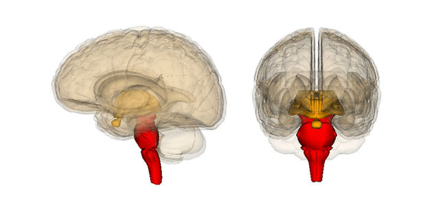
Your brainstem is a central axis that connects your cerebrum (Latin for "brain") to your spinal cord and cerebellum. The brainstem is divided into three parts from north-to-south: midbrain, pons, and medulla. Among many functions, the brainstem influences our sleep-wake cycle, automatic breathing, heart rates, states of arousal, etc.
If he were alive today, my dad would be over the moon to read the latest research from BIDMC. For the first time, this study homes in on a small “coma-specific” region of the brainstem—the rostral dorsolateral pontine tegmentum—that seems to drive consciousness through functional connectivity with two other cortical brain regions. One of these cortical regions is the left, ventral anterior insula (AI), the other is the pregenual anterior cingulate cortex (pACC). These three brain regions appear to work together as a triad to maintain consciousness.
Earlier this year, Boes received an award from the American Academy of Neurology for identifying a neural “consciousness network” that was affected by brain lesions on specific regions of the brainstem associated with cases of coma and vegetative states.
In an April 2016 statement to BIDMC, Boes said, “We are investigating what regions of the brainstem are most critical to consciousness. Knowing the anatomy of this system offers great potential for the development of targeted therapeutic stimulation strategies to improve recovery from coma and other devastating disorders of consciousness.”
For this research, Boes used brain network imaging to identify and map brain networks in 12 patients who had become comatose following a localized brainstem injury. Then, he mapped the location of the injury relative to 24 other patients with brainstem injuries that did not cause coma.
His results showed that any injury to a small area in the pons section of the brainstem (the rostral dorsolateral pontine tegmentum) is predictive of coma. As Boes explains, “We were excited to discover that this single tiny area is critical to consciousness. When it is damaged, almost every patient became comatose.”

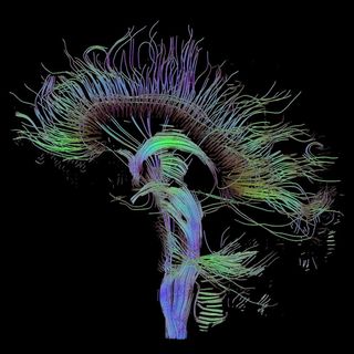
After pinpointing this tiny area of the brainstem as a potential “seat of human consciousness,” Boes and his colleagues mined data from the connectome project to identify other brain regions that were functionally connected to this particular region of the brainstem in healthy individuals. The Human Connectome Project (HCP) aided them in creating a detailed map of the neural network associated with consciousness. (To read more on HCP, check out my 2013 Psychology Today blog post, "What Is the Human Connectome Project? Why Should You Care?")
Lastly, the Harvard research team went back and examined fMRI brain images of 45 patients in a coma or vegetative state and discovered that activity in this novel brain network was selectively disrupted in patients lacking consciousness.
Arousal + Awareness = Consciousness
In conversations with my father about neurosurgery over the years, he would anecdotally describe dealing with the brainstem during an operation as being extremely sensitive (and potentially nerve-wracking) territory for him. As a brain surgeon, my father considered portions of the brainstem to be the most fragile and delicate parts of the human nervous system.
Through his extensive neurosurgical experience, my father knew that damage to the brainstem was often correlated with someone not waking up after surgery. That being said, I have no idea if he was consciously aware that the small rostral dorsolateral pontine tegmentum area of the brainstem was specifically what he colloquially referred to as “the spark plug of consciousness.”
In his November 2016 statement to BIDMC, David Fox, Director of the Laboratory for Brain Network Imaging and Modulation, summed up the latest findings of his team, "For the first time, we have found a connection between the brainstem region involved in arousal and regions involved in awareness, two prerequisites for consciousness. A lot of pieces of evidence all came together to point to this network playing a role in human consciousness."
The researchers point out that arousal combined with awareness are the two critical components of consciousness. Using this model, arousal is most likely regulated by the rostral dorsolateral pontine tegmentum in the brainstem.
The awareness aspect of consciousness may be directly linked to the connectivity of this brainstem region with the ventral anterior insula (AI) and pregenual anterior cingulate cortex (pACC). Although more research is needed, it appears that healthy functional connectivity of this brainstem-cortex network acts as a triad that may drive human consciousness.
Brainstem-Cortex Functional Connectivity Creates a "Consciousness Network"
In closing, Fox, who is also an Assistant Professor of Neurology at Harvard Medical School, concluded,
"We now have a great map of how the brain is wired up in the Human Connectome. We can look at not just the location of lesions, but also their connectivity. Over the past year, researchers in my lab have used this approach to understand visual and auditory hallucinations, impaired speech, and movement disorders. A collaborative team of neuroscientists and physicians had the insight and unique expertise needed to apply this approach to consciousness.
This is most relevant if we can use these networks as a target for brain stimulation for people with disorders of consciousness. If we zero in on the regions and network involved, can we someday wake someone up who is in a persistent vegetative state? That's the ultimate question."
The next step for this BIDMC team is to dive deeper into other data sets in which patients lost consciousness to clarify if the exact same brainstem-cortex network is always involved...or, if there are other brain regions that come into play regarding human consciousness. Stay tuned for more on this exciting topic!


