Dementia
Does the Cerebellum Fine-Tune Complex Cerebral Functions?
New research links atrophy of the cerebellum with Alzheimer’s and dementia.
Posted March 13, 2016

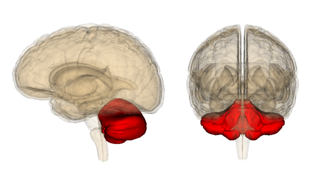
In 1504, Leonardo da Vinci made wax castings of the human brain and coined the term cerebellum (Latin for “little brain”). Historically, the cerebellum has been considered by most neuroscientists to be a region of the brain that is primarily involved in non-thinking activities such as coordinating the timing and precision of muscle movements.
Although it’s been five centuries since da Vinci made his iconic wax castings of the cerebellum, neuroscientists are just beginning to decode the mysterious “little brain.” This is a very exciting time to be researching and writing about the cerebellum. In recent years, a wide range of studies have shown, for the first time, that the cerebellum plays a pivotal role in many of our cognitive, emotional, and creative processes.

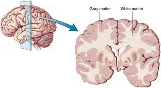
In March 2016, Ian Fyfe, MD, published an article, “Cerebellar Atrophy Has Disease-Specific Patterns," in the journal Nature Reviews Neurology. (Cerebellar means “of the cerebellum” and is the sister word to cerebral, which means “of the cerebrum.") In his review, Fyfe highlights two recent studies which have identified distinctive patterns of cerebellar atrophy that are related to wider patterns of brain network degeneration. Both studies found that the loss of gray matter brain volume in specific regions of the cerebellum is linked to Alzheimer's disease (AD) and frontotemporal dementia (FTD).
This research adds to a growing list of groundbreaking studies on the cerebellum that have been published in the past year. For example, an April 2015 study reported that adolescents with autism spectrum disorders (ASD) had abnormal cerebellar connectivity with areas of the cerebrum (Latin for "brain”) involved in higher-order cognitive functions such as decision-making, attention, and language. Other studies in the past year have linked ASD with atypical Purkinje cell development in the cerebellum.

In January 2016, a study identified a correlation between damage of cerebellar Purkinje cells in combat veterans who had experienced microblasts being strongly linked to treatment-resistant post-traumatic stress disorder (PTSD).
On the opposite end of the spectrum, in May of 2015 a study by researchers at Stanford University reported that robust and healthy cerebellar activity is linked to creative thinking and innovative breakthroughs.
To read more in-depth coverage of all the latest research on the cerebellum, check out the links to my previous Psychology Today posts at the bottom of this page.
In March of 2015, NPR's "Morning Edition" program did a feature story on the cerebellum that highlighted the work of Jeremy Schmahmann, M.D., from Massachusetts General Hospital.
After hearing the NPR piece, I emailed Dr. Schmahmann to introduce myself and ask him some questions about his groundbreaking research on the cerebellum. Through our correspondence, we realized that Schmahmann and my father—who spent many years as chief of neurosurgery at the Beth Israel Hospital in Boston—had a kismet "two degrees of separation" connection through Harvard Medical School.
Last spring, I also learned about Schmahmann's fascinating theory called “Dysmetria of Thought,” which is basically a hypothesis that the cerebellum helps to coordinate and fine-tune cognitive functions in the cerebral cortex much the same way it fine-tunes muscle movements by communicating with the left and right hemispheres of the cerebrum. (The left hemisphere of the cerebrum controls movements on the right side of the body; the right hemisphere of the cerebellum controls movements on the right side of the body, and vice versa.)
Below is a YouTube clip of Dr. Schmahmann's lecture, "The Cerebellar Affective Cognitive Syndrome: Implications for Neuropsychiatry," in which he explains his revolutionary theory of "Dysmetria of Thought" as related to the cerebellum and cerebral functions.
One of the key points of this video is the fact that, for eons, brain scientists have identified specific functions of various brain regions only after a stroke, accident, or some other malady damaged a specific area which triggered deficits or changes in personality. For example, Broca’s area (which is a brain area associated with speech) was discovered when Pierre Paul Broca identified impairments in two patients who had lost their ability to speak after an injury to the posterior inferior frontal gyrus area.
Therefore, in order to identify interventions that optimize brain function, neuroscientists rely first on making associations with various cognitive functions by identifying when and how things go wrong. For example, any discoveries about the cerebellum which pinpoint how the loss of brain volume (or functional connectivity) takes someone “south of zero” can be applied conversely to take someone “north of zero” by identifying lifestyle choices or pharmaceuticals that can fortify the structure and functional connectivity of the cerebellum in the opposite direction.
Future research on the cerebellum could lead to advances that help people with dementia, Alzheimer’s disease, autism, or PTSD lead better lives. This game-changing research on the cerebellum may also prove helpful for identifying actionable ways to optimize brain power and cerebral functions for people from all walks of life.
“Whatever the Cerebellum Is Doing, It’s Doing a Lot of It”
If you're wondering how I became interested in the cerebellum, here's the backstory. My father, Richard Bergland, was a neurosurgeon, neuroscientist, and author of The Fabric of Mind (Viking). My dad was obsessed with the cerebellum and passed this obsession on to me.
Although the cerebellum is only 10% of total brain volume, the cerebellar hemispheres hold well-over 50% of the brain’s total neurons. Based on the disproportionate number of neurons in the cerebellum, my father would often say, “We don’t know exactly what the cerebellum is doing, but whatever it’s doing, it’s doing a lot of it.”
In 2005, my father and I created a split- brain model that tagged the cerebrum as the "up brain" and the cerebellum as the "down brain." This was a direct and cogent response to the left brain-right brain model that my father had helped bring into the spotlight during the 1970s.
In addition to his own writing for a mainstream audience, my dad was also a medical expert for books such as Betty Edwards’ Drawing on the Right Side of the Brain. Although my father initially advocated for the left brain-right brain model, later in his life he became convinced that the most salient divide in the brain wasn't between the corpus callosum and the cerebral hemispheres, but rather between the midbrain which bridges the cerebrum and cerebellum, and the vermis.
When my father passed away suddenly of a heart attack in 2007, I made a vow that I would do my absolute best to finish his life’s work by keeping my antennae up for new research on the cerebellum. My goal is to connect-the-dots of the latest neuroscience in new and useful ways for a general reading audience. This daily commitment is why I am writing this blog post... Every day, I wake up hoping there will be new breakthroughs that advance our understanding of the mysterious and powerful cerebellum.
Below is a simple sketch I drew which maps how the various brain hemispheres might be interconnected in ways that fine-tune both our muscle movements and thoughts.

Atrophy of the Cerebellum Is Linked to Alzheimer’s Disease and Dementia
The past month has produced an abundance of fruitful new research regarding the connection between the cerebellum and complex cerebral functions. As I mentioned earlier, Ian Fyfe recently highlighted two different studies as breaking new ground by linking a loss of brain volume in the cerebellum as being associated with cognitive decline observed in both Alzheimer’s disease and Frontal Lobe Dementia.
Historically, scientists have focused solely on atrophy in the cerebral cortex as being associated with neurodegenerative diseases such as dementia. Until recently, the cerebellum has typically gone under the radar. However, the latest research suggests that atrophy patterns observed in the cerebellum may be an intrinsic part of a network-based degeneration framework in which neuropathology spreads across connectivity networks between all four brain hemispheres.
The first study, “Network-Selective Vulnerability of the Human Cerebellum to Alzheimer’s Disease and Frontotemporal Dementia,” was conducted by researchers in Australia and published in the February 2016 issue of the journal Brain. The researchers identified that specific neural networks between cerebellar circuits—which share extensive connections with the cerebral cortex—might be selectively targeted by certain neurodegenerative diseases.
For this study, neuroscientists examined the structural atrophy in the cerebellum across common types of neurodegenerative diseases, and characterized the functional connectivity patterns of these cerebellar atrophy regions.
The Australian researchers concluded that Alzheimer’s disease and frontotemporal dementia are associated with distinct and circumscribed atrophy in the cerebellum. This is the first time that researchers have demonstrated the selective vulnerability of the cerebellum to common neurodegenerative diseases. The researchers of the study concluded,
“Our work also has direct implications on the cerebellar contribution to the cognitive and affective processes that are compromised in neurodegeneration as well as the practice of using the cerebellum as reference region for ligand neuroimaging studies.”
The second study, “Patterns of Regional Cerebellar Atrophy in Genetic Frontotemporal Dementia,” was conducted by researchers at University College London (UCL) and published in the February 2016 issue of Neuroimage: Clinical.
Frontotemporal dementia (FTD) is a heterogeneous neurodegenerative disorder with a strong genetic component. The cerebellum hasn't traditionally been considered to be involved in FTD, but recent research suggests a potential link to cerebellar atrophy because the cerebellum is extensively connected to different brain regions in the cerebrum, including key areas involved in FTD.
The researchers point out that although the vermis (which connects the left and right hemisphere of the cerebellum) has long been known to be involved in the integration of sensory input regarding motor commands, “it is also considered the ‘limbic cerebellum’, involved in the modulation of emotions and social behaviors, based on its connections with the limbic brain structures.”
Is Gray and White Matter Integrity Between the Cerebrum and Cerebellum Interconnected?
Yesterday, I wrote a Psychology Today blog post, “Want to Bulk Up Your Brain? Burn Some Calories Via Exercise,” based on a new study which found that burning calories through physical activity was linked to increased gray matter volume in the cerebrum. Other recent studies have found that a lack of exercise is linked to reduced gray matter brain volumes in the cerebrum.

Along these lines, in an August 2015 study, researchers from the Beckman Institute at the University of Illinois at Urbana-Champaign reported that aerobic exercise and physical fitness improves the integrity of white matter tracts which connect the gray matter in various brain regions. This improvement in white matter integrity was associated with improved cognitive flexibility and spontaneous brain activity in older adults.
In The Athlete’s Way, I discuss that atrophy of the cerebellum is linked to sedentarism. People who are bedridden for more than six months can lose up to 23 percent of Purkinje cell volume in the cerebellum.
Based on the latest findings, one could make an educated guess that physical activity might stimulate gray matter growth via neurogenesis in both hemispheres of the cerebrum and cerebellum. Conversely, sitting all day could cause the gray matter volume in all four brain hemispheres to shrink while simultaneously reducing the functional connectivity of white matter between brain regions.

Albert Einstein said of E=mc2, “I thought of it while riding my bicycle.” Einstein was notorious for taking long walks around the Princeton campus, bicycling regularly, and being a virtuoso violin player by the age of seven. Interestingly, in 2013 a posthumous study of Albert Einstein’s cerebral brain hemispheres revealed that he had extraordinarily robust connectivity between the left and right hemispheres of his cerebrum that may have sparked his brilliance.
Although it’s pure speculation on my part, I wouldn’t be surprised to learn that Einstein also had robust structure and functional connectivity between his cerebellar hemispheres based on his physical activity and musicality.
One of the primary reasons I’m on a crusade to put the cerebellum in the spotlight (and make cerebellar a household word) is my belief that the key to maximizing each and every individual’s human potential lies in optimizing the structure and functional connectivity of all four brain hemispheres throughout his or her lifespan.
As the father of an 8-year-old, I’m fortunate that my daughter’s daily routines and education represent an equal blend of crystallized cerebral learning of “explicit” knowledge and cerebellar “implicit” learning. In addition to cerebral activities that flex her "book smarts," my daughter's weekly routine includes cerebellar activities such as tennis, swimming, ballet, taekwondo, piano and violin lessons, making art, etc. Collectively, these 'up brain-down brain' activities fortify the development and connectivity of her cerebellum and cerebrum.
Sitting Still and Memorizing Facts All Day Stagnates the Cerebellum
Unfortunately, with the overemphasis on standardized test scores, there is a knee-jerk reaction to cut funding for arts and athletic programs in public schools because most people don’t consider these programs to be related to academic achievement. I believe this is shortsighted for many reasons.
I am privileged enough to be a graduate of Hampshire College, which is one of the few colleges in the country with no tests or grades. At Hampshire, you don't ever have to memorize facts or feel pressured to get an A+ on an exam. I believe that taking the emphasis off crystallized intelligence and test scores leads to more fluid thinking.
The pedagogy of Hampshire College nurtures the connectivity between the cerebellum and the cerebrum. When someone is given the freedom to "unclamp" his or her prefrontal cortex and cerebral thinking, it fosters creativity by allowing the cerebellum to integrate and fine-tune complex ideas between all four brain hemispheres in new and unexpected ways. This type of thinking leads to Eureka! moments, patents, trademarks, etc.
I'm on a mission to collect as much science-based data that confirms the importance of daily activities that strengthen the architecture and connectivity of all four brain hemispheres from a young age in an attempt to persuade U.S. policymakers to fund arts and sports programs. In the long-run, our work force and economy will benefit by allocating funds for art, music, and athletic programs in American public schools.
As a nation, if we want the next generation of young Americans to have V12 levels of brain power, we need to take the emphasis off standardized testing and focus equally on developing every child’s cerebellum and cerebrum simultaneously. Strictly focusing on the crystallized knowledge held in the cerebrum—and forcing kids to sit still all day in school cramming their heads full of facts and figures—will backfire in the long-run if we neglect the cerebellum and allow it to atrophy.
In order to facilitate fluid intelligence and creative thinking, I believe it’s imperative for parents, educators, and coaches to have the resources that allow our children to stay physically active during recess, play sports, and express themselves through arts and music. Not only will this benefit the health of their bodies, it will benefit the structure and functional connectivity of all four hemispheres within their brains.
Conclusion: New Technology Will Help Decode the Mysterious Cerebellum
In closing, there was one other neuroscientific breakthrough this week that could open more doors in terms of our understanding of the cerebellum. Researchers at Max Planck Florida Institute for Neuroscience announced that they've fine-tuned revolutionary new techniques for assessing the motor learning activity of Purkinje neurons for days, weeks, or even months in mice while they're awake.
The March 2016 study, "Chronic Imaging of Movement-Related Purkinje Cell Calcium Activity in Awake Behaving Mice,’" was published in the Journal of Neurophysiology.
These state-of-the-art techniques will allow neuroscientists to pinpoint specific characterizations of Purkinje cells in real-time as a mouse engages in motor activity and other behaviors. In a press release, co-author Jason M. Christie, Ph.D., said, "Our work brings significant insight into the understanding of neural circuits within the cerebellum, which will be essential to understand how these circuits are altered in pathological conditions."
This is a very exciting time to be researching and reporting on the cerebellum. I bet that if Leonardo da Vinci were alive today, he’d be pleasantly surprised to know that the “little brain," which has been in the shadows for so long, is getting top billing in the 21st century. I know for sure that my father would be thrilled to see that advances in brain imaging technology are helping to solve the enigmatic riddle of what all those neurons in the cerebellum are actually doing ... Stay tuned for more cutting-edge research on the cerebellum.
To read more on this topic, check out my Psychology Today blog posts,
- "Einstein's Genius Linked to Well-Connected Brain Hemispheres"
- "Too Much Crystallized Thinking Lowers Fluid Intelligence"
- "The 'Right Brain' Is Not the Only Source of Creativity"
- "The Neuroscience of Madonna's Enduring Success"
- "Can Physical Activities Improve Fluid Intelligence?"
- "Musical Training Optimizes Brain Function"
- "The Neuroscience of Imagination"
- "The Cerebellum May be the Seat of Creativity"
- "Kids and Classrooms: Why Environment Matters"
- "Childhood Creativity Leads to Innovation in Adulthood"
- "Why Does Overthinking Sabotage the Creative Process"
- "Superfluidity: Decoding the Enigma of Cognitive Flexibility"
- "How Are Purkinje Cells in the Cerebellum Linked to Autism?"
- "Cerebellum Damage May be the Rood of PTSD in Combat Veterans"
© 2016 Christopher Bergland. All rights reserved.
The Athlete’s Way ® is a registered trademark of Christopher Bergland.
Follow me on Twitter @ckbergland for updates on The Athlete’s Way blog posts.


