Brain imaging, using methods like Magnetic Resonance Imaging (MRI), electroencephalograph (EEG), and many others, is an important research area for ADHD. It’s also a “hot topic” with periodic excited claims in the media. (I discuss this in the context of various new treatment claims for ADHD and try to separate the wheat from the chaff in my book, Getting Ahead of ADHD). The bottom line: brain imaging cannot diagnose ADHD or its subtypes.
What are these methods? The most common brain research tool in children is probably the MRI. Some of you have had an MRI for medical purposes. The child lies on a movable table and slides into a large metal cylinder or doughnut. The cylinder houses a large magnet that uses magnetic pulses to detect changes in the flow of oxygenated molecules.
In turn, that is used to infer blood flow, and thus infer neural activity. (No radiation is involved). It can show how different parts of the brain relate or communicate during a task or at rest. These communication patterns have characteristic structures and forms. MRI can also show the brain structure—how large or thick are different parts of the brain.
EEG uses electrodes (sensors) on the scalp to detect electrical signals from the brain. All neural activity is electrochemical. Therefore, variations in electrical waves, while slight by our usual standards of volts and currents, are measurable with these sensitive electrodes. (They do not transmit any electric current.)
EEG is very time-sensitive (detecting changes in electrical activity down to 1 millisecond). That’s how we know, for example, that it takes about 100 milliseconds for the average adult to consciously “see” something they just saw. Brain waves oscillate or weave in regularized patterns. The method also measures these.
Many other methods are also used. SPECT measures the flow of an injected radioactive tracer through the blood to map the brain (it is a favorite of the popular writer Daniel Amen). There are also MEG, fNIRS, PET (similar to SPECT), and others.
In all methods, the idea is to detect how the brain differs in people with ADHD from other people, or how sub-groups with ADHD may be unique. Some studies also try to “diagnose” ADHD by running different mathematical models on the scans.
My research team and others are now undertaking a new generation of studies using advanced nonlinear equations (called machine learning) to improve prediction. These are powerful methods, and there is reason to be hopeful.
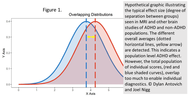
However, results from existing studies do not yet offer clinical value. One limitation is that sample sizes tend to be extremely small (often less than 100 children)—such samples are prone to chance findings that will not generalize, no matter how clever the analysis is. Scientists attempt to overcome this in various ways. However, the acid test is whether the prediction holds up in a completely new, independent sample of children. Typically, that check is not even done. When it has been done, results are generally disappointing. This is because the differences between ADHD and non-ADHD individuals are typically very small. They are big enough to see a group difference, but not big enough to reliably pick out which group an individual is in. (See Figure 1).

An analogy would be the differences in height between teenage boys and girls. While a group average would show that boys are taller than girls, it’s not possible to predict whether someone is a boy or girl by knowing their height.
Instead, we would try to combine many measures that each give a little different information. The idea in brain imaging is to use many different points of measurement. However, this hasn’t yet been successful at predicting accurately enough to be useful in a clinic.
This generalizability problem is challenging. It will be some time before it is solved. However, many research groups, our own included, are trying hard to do this. We have high hopes that we will eventually succeed in identifying important subgroups of children with ADHD based on variations in brain development.
To this point, any clinical insights “gleaned” from a brain scan can also be obtained by a standard expert clinical evaluation.
So stay tuned. Future findings should change the picture for the better with improved tools and methods. We are among the groups working on that. But for now, remain skeptical of claims for breakthrough brain imaging diagnostics for ADHD. This includes FDA-approved EEG and other measures, which are approved because they are safe, but not because they have demonstrated accuracy.
I advise against seeking brain imaging for a clinical case of ADHD (in the absence of other medical indications, like a head injury or seizure). Nothing has changed with recent publications or press reports. Save your hard-earned dollars for a qualified clinical evaluation and care.
We will be tracking this literature, and I will share significant findings with you.
As always, let’s keep our eye on the science for reliable answers.
Please note: Dr. Nigg cannot advise on individual cases for ethical, legal, and logistical reasons.


