Fear
Subconscious Fear Exposure Helps Reduce Phobias, Study Finds
Subliminal exposure to spider images reduces fear response in arachnophobes.
Posted February 9, 2017

The American Psychiatric Association estimates that approximately one in ten people in the United States suffer from some type of phobia. About 40 percent of phobias are related to creatures such as spiders, snakes, rats, lizards, bats, etc. If you are among the millions of people who are spider-phobic (arachnophobia) or have an abnormal fear of other vermin, there is good news.
A new study offers a potentially revolutionary treatment option for anyone suffering from an atypical fear of spiders or other phobias. For arachnophobes, the researchers found that subconscious exposure to a spider image (such as the tarantula above) for a millisecond—without any conscious awareness of viewing the image—was more effective at reducing a fear of spiders than longer, conscious exposure. The February 2017 findings were published in the journal Human Brain Mapping.
Although phobias are often considered to be an irrational fear, most of the stimuli that trigger phobic responses have deep roots in our evolutionary biology that stem from a justifiably hardwired fear of anything that could have threatened our individual or collective survival as a species. Interestingly, humans are born with a host of innate fears that are part of our neurobiology from birth but reside below the threshold of conscious awareness.
Humans respond to any fearful stimuli via an interplay between subcortical ("non-thinking") brain regions and cerebral ("thinking") cortical brain regions such as the frontal cortex. For decades, I've been researching the hypothesis that implicit learning and fear-based conditioning or avoidance behaviors are driven by subcortical brain regions seated below the conscious awareness of cortical regions in the cerebral cortex. The latest research on backward masking adds valuable insights to this hypothesis.
As an example of subconscious fear responses, anyone who has ever mistaken a harmless piece of rubber on a path or in your backyard for a snake knows how deeply embedded a fear of serpents is seared into your subcortical brain regions. This primal subcortical fear of snakes is why your body will automatically jump away at the sight of an innocuous garden hose in the yard before your conscious mind and cortical brain regions have time to rationalize or decipher that the garden hose poses no danger.
The Dynamic Interplay Between the Frontal Cortex and Caudate Nucleus Influences Fear Responses
For the new study on arachnophobia, a team of researchers including Bradley Peterson, director of the Institute for the Developing Mind at Children's Hospital Los Angeles, and Paul Siegel, associate professor of psychology at Purchase College of the State University of New York, used fMRI brain imaging and a technique called "backward masking" to pinpoint brain areas involved in conscious and subconscious fear processing. (Very brief exposure to potentially phobic stimuli followed by longer exposure to a non-threatening "masking" image that distracts from cognitive awareness of the threatening image is called "backward masking.")
To test neural activity during very brief subconscious vs. longer conscious exposure to phobic stimuli, the researchers recruited a group of 21 spider-phobic study participants and a cohort of 21 people who weren't afraid of spiders. All 42 participants were exposed to three conditions: (1) Very brief exposure (VBE) to masked images of spiders, severely limited awareness; (2) clearly visible exposure (CVE) to spider images, full awareness; and (3) masked images of flowers (control).
Then, Peterson and colleagues examined the degree to which specific areas of the brain involved in fear processing and deciding how to respond to a phobic image were activated when someone was consciously aware or unaware of the spider image. Interestingly, they found that even though non-conscious awareness didn't register in a way that could be brought to mind or was cognizant, this type of exposure caused subliminal fear responses to skyrocket.
Surprisingly, the amygdala (which is widely considered the hub of fear responses and processing) wasn't the focus of this study. Instead, the fMRI neuroimaging honed in on the activity of the caudate nucleus—which regulates emotional fear responses and works with the frontal cortex to figure out how to respond to threatening stimuli. As you can see by this colorful fMRI image, both the caudate and frontal cortex lit up more significantly during subconscious exposure to phobic images when someone was "unaware" of a spider image.
Subcortical Structures Are Taking Center Stage in the Early 21st Century
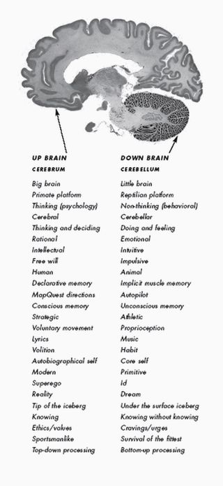
The caudate nucleus is a small subcortical brain structure in the dorsal striatum, which is housed in the basal ganglia. In recent years, there's been growing interest among neuroscientists to identify how subcortical “non-thinking” regions such as the basal ganglia, brainstem, and cerebellum (Latin for "little brain") interact with cortical “thinking” brain regions such as the frontal cortex, which is housed in the cerebrum (Latin for "brain").

In 2005, my father and I created the "Bergland split-brain model" as the foundation for The Athlete's Way program—which relies on taking a two-pronged approach for optimizing your mindset and maximizing performance based on implicit (subcortical) and explicit (cortical) learning and memory. My father, Richard Bergland, was a neuroscientist, neurosurgeon, and author of The Fabric of Mind. Tragically, he died unexpectedly a few weeks after our split-brain model was published by St. Martin's press in 2007 and didn't live to witness 21st-century advances in neuroscientific research catch up to his visionary hypotheses about how the brain works.
In the late 20th century, my dad had an "aha!" moment when he realized that the dynamic interplay between subcortical and cortical brain regions needed to be put in the spotlight. The Athlete's Way became a vehicle for my father (and me) to deliver potentially radical neuroscientific ideas to a general audience that were too iconoclastic at the time to be published in peer-reviewed journals.
The Bergland split-brain model refers to cortical regions as "up brain" and uses the terminology "down brain" to describe subcortical regions. "Up brain-down brain" is a direct and cogent response to the ubiquitous but deeply flawed "left brain-right brain" dyadic model. As I describe on p. 25 of The Athlete's Way,
"The salient divide in the brain is not from east to west or from right to left. Instead, it is from north to south. The down brain is our emotional and intuitive center and may even hold our personal and collective unconscious mind. It stores all [evolutionary] long-term memories, an ancient primal defense mechanism."
If you'd like to read more on this topic, in a January 2017 Psychology Today blog post, "Radical New Discoveries Are Turning Neuroscience Upside Down," I give an overview of a broad range of cutting-edge research that gives top billing to the cognitive and behavioral power of previously underestimated subcortical brain regions buried beneath the "thinking cap" of the cerebral cortex.
Because many of the ideas presented in The Athlete's Way about subcortical brain regions were ahead of their time a decade ago, I've kept my antennae up for 21st-century technological advances in neuroimaging that provide state-of-the-art empirical evidence which corroborates many of the educated guesses my father made decades ago and that I first published in 2007.
Needless to say, I was thrilled to read about the new research by Peterson et al. because it advances our understanding of the dynamic interplay between subconscious/subcortical brain regions and conscious/cortical brain regions. In a statement, first author of the recent arachnophobia research using backward masking, Paul Siegel described his team's findings,
"Counter-intuitively, our study showed that the brain is better able to process feared stimuli when they are presented without conscious awareness. Our findings suggest that phobic people may be better prepared to face their fears if, at first, they are not consciously aware that they've faced them."
Bradley Peterson added, "Although we—expected and observed—activation of the neural regions that process fear, we also found activation in regions that regulate the emotional and behavioral responses to fear—reducing the conscious experience of fear."
Current therapies to cope with phobias often involve directly confronting the feared stimulus to create desensitization. Unfortunately, consciously facing fears can cause young people to experience significant emotional distress and may actually be less effective than starting the process with subconscious backward masking. Peterson is in the process of fine-tuning potential ways to use backward masking to treat children and adolescents with a variety anxiety disorders and phobias.
Backward Masking Can Fortify Self-Belief and Athletic Performance
In 2014, there were two studies that reported on the power of backward masking and subconscious messaging to improve athletic performance and minimize negative self-beliefs about getting older. I reported on this research in a Psychology Today blog post, "Subliminal Messaging Can Fortify Inner Strength." This previous research dovetails with the new 2017 findings of using backward masking to help overcome phobias.
The first example of using backward masking to improve performance is a December 2014 study, “Non-Conscious Visual Cues Related to Affect and Action Alter Perception of Effort and Endurance Performance,” conducted by Professor Samuele Marcora at the University of Kent in collaboration with colleagues at Bangor University and published in the journal Frontiers in Human Neuroscience.
In this experiment, the researchers flashed subliminal cues, such as action-related words or happy vs. sad faces on a digital screen while endurance athletes were exercising on a stationary bicycle.
The subliminal words and faces appeared on a digital screen for less than 0.02 seconds and were masked by other visual stimuli making them unidentifiable to the participant's conscious mind. When the athletes were presented with positive visual cues like "go" and "energy" or were shown happy faces they were able to exercise significantly longer compared to those who were shown sad faces or words linked to inaction or fatigue.
Another example of the power of subliminal messaging and backward masking was conducted at Yale University. The October 2014 study, “Subliminal Strengthening: Improving Older Individuals, Physical Function Over Time With an Implicit-Age-Stereotype Intervention,” was published in the journal Psychological Science.
In this study, the researchers used backward masking to examine whether exposure to positive age stereotypes could weaken negative age stereotypes and lead to more vitality and healthier outcomes.
The Yale School of Public Health researchers found that older individuals who were subliminally exposed to positive visual cues and stereotypes about aging showed improved physical functioning that lasted for several weeks.
In this study, some of the participants were subjected to positive age stereotypes on a computer screen that flashed words such as "spry" and "creative" at speeds that were too fast to be picked up consciously. This was the first study to show that backward masking can improve attitudes aging and physical function among senior citizens.
Stay tuned for future research on specific ways that backward masking can be used to overcome fear and phobias. Hopefully, future research will also fine-tune ways to use backward masking and implicit learning to improve confidence, physical performance, and someone's overall capacity to seize the day.
References
Paul Siegel, Richard Warren, Zhishun Wang, Jie Yang, Don Cohen, Jason F. Anderson, Lilly Murray, Bradley S. Peterson. Less is more: Neural activity during very brief and clearly visible exposure to phobic stimuli. Human Brain Mapping, 2017; DOI: 10.1002/hbm.23533
B. R. Levy, C. Pilver, P. H. Chung, M. D. Slade. Subliminal Strengthening: Improving Older Individuals' Physical Function Over Time With an Implicit-Age-Stereotype Intervention. Psychological Science, 2014; DOI: 10.1177/0956797614551970
B. R. Levy, C. Pilver, P. H. Chung, M. D. Slade. Subliminal Strengthening: Improving Older Individuals' Physical Function Over Time With an Implicit-Age-Stereotype Intervention. Psychological Science, 2014; DOI: 10.1177/0956797614551970


