Neuroscience
Have Scientists Found the Brain's "Engine"?
Neuroscience research shows this part of the brain sets the global stage.
Posted February 3, 2018 Reviewed by Hara Estroff Marano
Scarecrow: I haven't got a brain... only straw.
Dorothy: How can you talk if you haven't got a brain?
Scarecrow: I don't know... But some people without brains do an awful lot of talking... don't they?
Dorothy: Yes, I guess you're right.―L. Frank Baum
What makes the brain tick? What keeps the constant stream of energy welling up within ourselves when we are alert and awake? What is the brain’s “engine room”, producing the momentum which makes everything else go?
This is an important question because the brain is always in motion. There is always an underlying level of brainwave activity, which means that neurons, even in a state of complete rest, are oscillating. It means that in health, the brain is always doing something. The brain is not like a computer, in this sense. It never truly turns off or shuts down, unless something is very seriously wrong.
Researchers now report that the deepest, oldest part of the brain sets the level of activation for the newest part of the brain, the cerebral cortex. Knowing how the brain is engineered paves the way for novel approaches to disorders such as Alzheimer's disease and depression.
Understanding what keeps the brain going, what provides the energy that drives the whole system, is of obvious value for understanding health and illness and for developing tools to alter brain activity in more predictable ways. People talk about the default mode network (DMN), which generates what our state of mind is like when we are awake and at rest, what thoughts drift in and out, what the emotional tone is like, and so on. But what keeps the DMN going, from down below?
Understanding, for example, that areas of the surface cortex can inhibit excessive emotion generated by the hard-to-reach limbic system enables use of a strong pulsing magnetic field to buff up cortical activity in a specific area and make it easier for people with depression to change their mood for the better and to deal with repetitive, negative thoughts. Use of such a device is called "Transcranial Magnetic Stimulation", and it is now considered a standard psychiatric treatment.
The deep, old parts of the brain, located just above the spinal column and in the center of the brain, collectively known as the basal forebrain, are evolutionarily ancient structures shared by humans with other animals with the same basic brain blueprint.
The basal forebrain is implicated in basic brain processes, sending bundles of neurons called “projections” to many other brain areas; some are scattered throughout the whole cortex. The cortex is the outermost surface of the brain, more developed in more intelligent animals. Humans additionally have a “neocortex”, a wafer-thin film of tissue that enhances our mental function in comparison with that of other animals. The basal forebrain also sends projections to subcortical (“under the cortex”) areas of the brain, including the amygdala, involved with strong emotions including fear, and the hippocampus, involved with memory and context, as well as other key areas.
Signals in the brain are carried in many ways, mainly electrically and chemically. These chemicals are myriad, a complex brew of small molecules, short proteins (“neuropeptides”) and a variety of hormonal molecules, often generally referred to as “neurotransmitters”. Many of these molecules are shared by the nervous, endocrine and immunological systems, while some are localized within the brain.
When it comes to the basal forebrain, the projections it sends are associated with several major neurotransmitters with different general effects, such as glutamate and excitation, GABA (gamma-aminobutyric acid) and inhibition, and acetylcholine (abbreviated "ACh" and referred to as "cholinergic") with important regulatory effects. ACh, for instance, can decrease activity in the cortex by slowing down synchronization of neurons, and is implicated in Alzheimer’s disease. Cholinergic projections from parts of the basal forebrain strongly influence level of arousal (e.g. alertness and drowsiness) and thinking.
Researchers have been interested in a particular part of the basal forebrain, called the “nucleus basalis of Meynert” (NBM), as a potential driver of resting-state brain networks such as the DMN. While brain networks generate local activity through interactions among the parts making up those networks, they are also dependent upon the background global level of brain activity to stay afloat. It’s a little bit like keeping your muscles slightly tensed but loose enough to move quickly when waiting for a tennis serve or a ball to come over the plate; there’s a baseline tone upon which responsiveness is optimized, creating a nimble ("complexly adaptive") system.
Does the NBM play a role in pushing up this base level of global brain tone? In order to investigate this question, Turchi and colleagues (2018) inactivated and reactivated tiny areas of the NBM in macaque monkeys, measuring their cerebral cortex via functional magnetic resonance imaging (fMRI). Because the NBM has specific subregions, separated by mere millimeters, researchers hoped to learn how specific the NBM was at driving activity in the cortex.
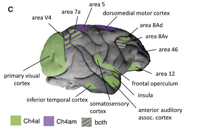
They found that when they switched deep forebrain subregions on and off, very selective areas of the cortex also switched on and off. This diagram shows how precise the bottom-up influence is; the purple and green areas of the cortex correspond to tiny clusters of cell activity in the forebrain:

While there is some overlap, and perhaps some areas that are not covered by these parts of the NBM, this study shows evidence of a one-to-one mapping of areas of the basal forebrain, an evolutionarily ancient area of the brain, onto the more recently developed cerebral cortex. These areas all have specific functions, and are embedded as part of various higher-order brain networks. The forebrain, leading from behind, as it were, sets the baseline levels of activation for whatever specific brain regions are doing.
What about brain networks? Does the forebrain selectively influence the key functional networks, including the default mode network and others, such as the salience network, which determines what we pick up on and what we ignore around us, and the executive control network, which guides motivation and attention and helps regulate emotional states? Prior research has shown that the amygdala (often thought of only for fear but involved intense positive and negative experiences) switches activity in specific brain networks, so what about the NBM?
As it turns out, in this study scientists did not see a connection between specific network activity and switching areas in the forebrain. While overall tone is influenced via cortical and subcortical areas projected to by the forebrain, so far a specific connection from the forebrain onto common networks has not been identified. Yet, it may be via regions that have not yet been tested.
The research is important from a basic neuroscience perspective as well as from clinical and neuroengineering perspectives. Understanding that areas such as the forebrain, highly conserved across species, set the activation tone for the cerebral cortex moves basic knowledge a significant step along. It is likely, but needs to be studied, that what is observed in primates is similar to what is happening in us humans. And of course, there are many brain areas that project onto and influence activity in the forebrain itself...
For conditions of disordered arousal, such as dementia, and other neuropsychiatric conditions, or for understanding how medical illnesses may affect the brain as well, mapping out the function of the forebrain is critical for developing therapeutic targets. For example, it could be possible to insert a brain-stimulating electrode into key areas of the forebrain to amp up global cortical activity in patients with decreased function, or even to use noninvasive neuromodulation techniques such as transcranial direct current stimulation to reach deeper brain areas.
Techiques like deep brain stimulation are already being used to treat Parkinson's disease and severe major depressive disorder, and severe obsessive compulsive disorder can be alleviated by destroying tiny amounts of brain in very specific areas. If we know how the brain is engineered, we can develop and test targeted neuromodulation approaches based on that understanding.
Research is being conducted to continue refining understanding of how the brain works, how networks of the brain function in relation to anatomical areas and physiological processes, and how basic neuroscience can be translated into clinical applications—and possibly human performance enhancement.
References
Turchi et al., The Basal Forebrain Regulates Global Resting-State fMRI Fluctuations, Neuron (2018), https://doi.org/ 10.1016/j.neuron.2018.01.032


