Neuroscience
What Clinicians Can Learn From Our "Brain Fingerprints"
How we use our neural networks is part of what makes us all unique.
Posted June 28, 2022 Reviewed by Ekua Hagan
Key points
- Patterns in neural networks are both similar across people and unique to individuals.
- A study has revealed that those suffering from complicated grief have a specific connection pattern in the brain that can be shown on an fMRI.
- Identifying differences in neural network patterns can help clinicians develop more effective treatments for individuals.
One of the most useful tools in the neuroscientist’s arsenal is the functional magnetic resonance image or fMRI. fMRIs allow researchers to watch the brain work as we do everything from solving complex problems to responding to a flash of light. fMRIs measure changes in blood flow that happen when cells in the brain go to work.
If you’re having an MRI scan done on your functioning brain, you will be placed in a large tube with magnets that spin around you creating a magnetic field. Those magnets make hydrogen atoms in the blood supply spin and a computer translates different rates of spin (different amounts of oxygen) into a color-coded image indicating levels of activity in the brain.
You are often given a mental task to perform while in the MRI machine, although you can also scan the resting brain and watch what neurons are doing in a resting-state fMRI (rsfMRI). Specific regions of the brain that are used during this task work harder than do other areas and so require more blood supply and more oxygen. They show up coded in a bright red color in the scan. Cooler colors like blues and greens indicate less activity in a region.
What is a connectome?
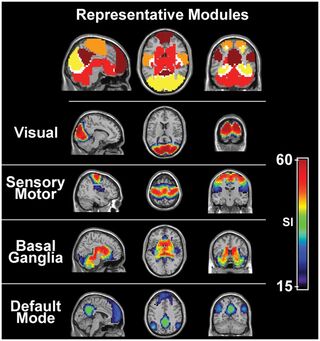
If you scan the whole brain, you can correlate synchronous activity in different anatomically separate areas of the brain and develop a map of the way that brain regions work together, called a connectome.

There are at least seven main networks in the human brain (the exact number depends on how you define a network). A word of caution here—there is both agreement and disagreement about just what exactly these networks are and how they are organized; this is just one description.
There is the default mode network (consisting of those parts of the brain that are active when we’re not really doing anything specific, when we’re just contemplating things), the sensory-motor network (used when we’re performing a motor task or when we’re coordinating our movements for that task), the visual network (active when we’re using visual information to solve problems, recognize patterns and faces, identifying objects and analyzing the movement of objects in the visual field), the central executive network (used to integrate information from other networks and working memory in making decisions), the limbic network (involved in determining our reaction to a stimulus, as well as the emotion associated with that reaction, reward and motivation), the salience network (determining what is most important in the world around us and moderating how the other networks, in particular the default and central executive networks will react) and the dorsal attention network which guides our attention to what is most important or most active in the world around us.
One of the most interesting revelations emerging from this research is that, despite the similarities in these networks in all human beings, the ways that we each use these networks vary from person to person. We tend not only to use these networks in a way that differs from others but is also consistently typical of “us” across time. The ways different brain networks are used by each of us is so individual and unique that they can be used to identify us from all others, like a brain fingerprint, just from looking at a scan.
Researchers and clinicians are interested in these individual patterns of use because they may help design more effective treatments for individuals who are experiencing pain, distress, or difficulty.
Connectomes and recovery from grief
Grief over the loss of a loved one is one example of how connectomes might help people struggling with loss. Grief is awful and loss is painful, but for most people, it is blessedly transient, eventually resolving over time. However, some individuals (some 7-10%) experience complicated grief—a grieving response that is more intense, protracted, and debilitating than the typical reaction to loss.
A study done by Chen, et al., in 2020 used rsfMRI to examine the activity in the limbic network in grieving people who had lost their loved one within the past year. In particular, they were interested in the connection patterns stemming from a part of the emotion circuit called the amygdala.
They found that the amygdala was more active in grieving individuals who were suffering from complicated grief than in healthy controls. In addition, activity between the amygdala and the rest of the limbic network, the default network, and both the executive control and salience networks tended to increase as the grieving symptoms worsened over time.
Their conclusion was that this kind of fMRI scan might help clinicians determine who was likely to experience complicated grief and allow early intervention for those individuals.
References
Description of networks is from Omniscient Neurotechnology at academy.o8t.com/
Chen, G., Ward, B.D., Claesges, S.A., Li, S-J. and. Goveas, J.S. (2020). Amygdala functional connectivity features in grief: A pilot longitudinal study. American Journal of Geriatric Psychiatry, 28(10), 1089-1101. doi:10.1016/j.jagp.2020.02.014.


