Addiction
Stanford Scientists Discover Surprising Cerebellum Functions
Cognitive process in cerebellar granule cells encodes the expectation of reward.
Posted March 21, 2017

In a serendipitous discovery, neuroscientists at Stanford University recently stumbled on previously unknown cognitive functions of the cerebellum. In a series of complex mice experiments using state-of-the-art brain imaging technology, the Stanford researchers found that specific neurons (granule cells) within the cerebellum learn and respond to anticipated rewards, or the lack thereof.
The new study from the Stanford Neuroscience Institute, "Cerebellar Granule Cells Encode the Expectation of Reward," was published March 20 online ahead of print in the journal Nature. (Cerebellar is the sister word to cerebral and means "relating to or located in the cerebellum.")
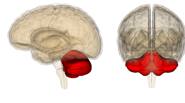
In 1504, Leonardo da Vinci made wax castings of the human brain and coined the term "cerebellum" (Latin for “little brain”) after identifying two small brain hemispheres tucked neatly under the relatively humongous left-right hemispheres of the "cerebrum" (Latin for "brain").

For centuries, neuroscientists considered the cerebellum to be the seat of “non-thinking” activities such as coordinating and fine-tuning muscle movements.
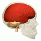
Until recently, both hemispheres of the cerebrum—and cortical regions in the “thinking cap” of the cerebral cortex—were considered the sole domain of cognitive processes. This is beginning to change. In recent years, a wide range of studies have begun to show (for the first time) that the cerebellum plays a mysterious but significant role in many cognitive brain functions.
For example, in February 2017, an international team of researchers reported that the cerebellum may play a previously unforeseen role in brain alterations associated with the addictive consumption of drugs.
These reports linking the cerebellum and drug addiction were based on a broad range of groundbreaking research published over the past two years. These cerebellar findings were compiled and featured in two different journals: Neuroscience & Biobehavioral Reviews and the Journal of Neuroscience.
In describing the objective of the first review, “Have we been ignoring the elephant in the room? Seven arguments for considering the cerebellum as part of addiction circuitry,“ the scientists, led by Marta Miquel Salgado-Araujo of UJI in Spain, describe their mission stating, “Our goal is not to review animal and human studies exhaustively but to support the inclusion of cerebellar alterations as a part of the physiopathology of addiction disorder.”
New Technology and an Accidental Discovery Waiting to Happen: Cerebellar Granule Cells Play a Cognitive Role in Reward Processing

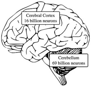
The March 2017 report from Stanford on the link between the cerebellum and encoding the expectation of reward dovetails with the aforementioned research on the cerebellum being part of addiction circuitry. The new Stanford research also advances our understanding of the mysterious neuronal power held in our brain's 60 billion granule cells. (Although the cerebellum is only 10 percent of brain volume, it houses up to 80 percent of your brain's total neurons, most of which are granule cells.)
One of the most noteworthy aspects of the new Stanford study is that the researchers accidentally stumbled on their potentially earth-shattering discovery that granule cells learn and respond to anticipated rewards. In some ways, the fact that the researchers didn’t design an experiment with the intent of proving a hypothesis adds an extra level of credibility to this first-of-its-kind discovery.
Mark Wagner, a postdoctoral fellow at Stanford led this research with Tony Kim, a graduate student in the lab of Mark Schnitzer, associate professor of biology and applied physics.
Schnitzer has a knack for developing unique methods for recording the brain activity in fruit flies, mice, and other living animals. One proprietary method Schnitzer recently developed, called "two-photon calcium imaging," offered the finite resolution that Wagner needed to study microscopic granule cells while mice were in motion.
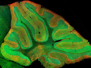
Because granule cells are packed so densely within the cerebellum, conventional techniques for recording granule cell activity don’t work very well, which has left neuroscientists with an incomplete picture of what the cerebellum is actually doing, until now.

My late father, Richard Bergland, was a 20th-century neuroscientist, neurosurgeon, and author of The Fabric of Mind (Viking). He was fascinated with the disproportionate distribution of neurons in the cerebellum. That said, my dad grew frustrated by technological limitations of his generation that made it impossible for him to empirically prove his 'educated guesses' about what the cerebellum was doing in his laboratory.
Like a broken record, he would say, "We don't know exactly what the cerebellum is doing. But whatever it's doing, it's doing a lot of it." If my father were alive today, I know he'd be over the moon to see that the revolutionary two-photon calcium imaging technique developed by Schnitzer is finally allowing Stanford neuroscientists to view granule cell activity in real time.
When Wagner began his research using two-photon calcium imaging, he was simply interested in monitoring and recording granule cells in real time as part of basic motor control functions of the cerebellum.
In order to study motor control, Wagner and his team needed to motivate their lab mice to move in the first place. So, they conditioned a reward-seeking behavior which was receiving a dose of sugar water after pushing a dispenser lever. While the mouse was pushing the lever and receiving a reward, Wagner and his team recorded granule cell activity in each mouse’s cerebellum.
Wagner was only expecting to find that granule cell activity was related to planning and executing physical movements. But in a Eureka! moment, Wagner observed that only some granule cells fired when the mouse pushed the lever for a reward. Surprisingly, other granule cells fired when a mouse was waiting for his or her sugary reward. And, yet another subset of granule cells fired when Wagner sneakily took away the anticipated Pavlovian rewards. The scientists write in the Nature abstract of their study:
"Tracking the same granule cells over several days of learning revealed that cells with reward-anticipating responses emerged from those that responded at the start of learning to reward delivery, whereas reward-omission responses grew stronger as learning progressed. The discovery of predictive, non-sensorimotor encoding in granule cells is a major departure from the current understanding of these neurons and markedly enriches the contextual information available to postsynaptic Purkinje cells, with important implications for cognitive processing in the cerebellum."
In a statement to Stanford, co-author Liquin Luo said, “It was actually a side observation, that, wow, they actually respond to reward." Wagner added, “we just didn’t know,” because historically the assumption was that granule cells only perform the most basic motor functions and no one had the tools to look closely at granule cells in action.
Moving forward, Wagner and his colleagues at Stanford are optimistic that this discovery could lead to something much bigger. In conclusion, Wagner said,
“Given what a large fraction of neurons reside in the cerebellum, there’s been relatively little progress made in integrating the cerebellum into the bigger picture of how the brain is solving tasks, and a large part of that disconnect has been this assumption that the cerebellum can only be involved in motor tasks. I hope that this allows us to unify it with studies of more popular brain regions like the cerebral cortex, and we can put them together.”
Stay tuned for more cutting-edge research on granule cells and the cerebellum. In the meantime, if you’d like to read my previous Psychology Today blog posts about the cerebellum click on this link.
References
Mark J. Wagner, Tony Hyun Kim, Joan Savall, Mark J. Schnitzer & Liqun Luo
Cerebellar granule cells encode the expectation of reward. Nature (2017) DOI: 10.1038/nature21726
Marta Miquel, Dolores Vazquez-Sanroman, María Carbo-Gas, Isis Gil-Miravet, Carla Sanchis-Segura, Daniela Carulli, Jorge Manzo, Genaro A. Coria-Avila. Have we been ignoring the elephant in the room? Seven arguments for considering the cerebellum as part of addiction circuitry. Neuroscience & Biobehavioral Reviews, 2016; 60: 1 DOI: 10.1016/j.neubiorev.2015.11.005
Barbara A. Sorg, Sabina Berretta, Jordan M. Blacktop, James W. Fawcett, Hiroshi Kitagawa, Jessica C.F. Kwok, Marta Miquel. Casting a Wide Net: Role of Perineuronal Nets in Neural Plasticity. The Journal of Neuroscience, 2016; 36 (45): 11459 DOI: 10.1523/JNEUROSCI.2351-16.2016


