Dementia
Using the Retina as a Biomarker for Brain Function
Retinal structure may help predict brain disorders.
Updated September 26, 2023 Reviewed by Abigail Fagan
Key points
- Thickness of the layers of cells that make up the retina reliably predict brain function.
- Thickness of the retinal nerve fiber layer has been linked to disorders like Alzheimer's disease.
- Retinal thickness could be a signal that patients might be at greater risk of developing Alzheimer's disease.
There’s an old saying, attributed to writers from Cicero to Shakespeare, that says the eyes are the windows to the soul. But it may be more accurate to say that your eyes, and in particular your retinas, are the windows into the functions of your brain.
Neuroscientists consider the retina and the optic nerve that carries information into the brain to be a part of the central nervous system, rather than a part of the sensory machinery of the eye. It makes sense then that we might be able to discern something about brain function from this relatively easily accessed part of it.

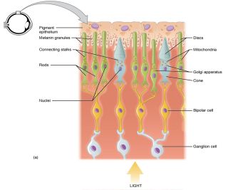
If you were to ride on a beam of light entering the eye, you would encounter three main layers of cells when you hit the retina which coats the back surface of the eyeball. The first layer, closest to the incoming light is made up of ganglion cells (GC). Slightly deeper, in the middle layer of the retina are the bipolar cells. Both GC and bipolar cells do not respond to the incoming light itself; instead, they are part of the chain of cells that carries the signal about light out of the retina and into the brain. The cells that actually create a signal when exposed to light are deeper still, at the very deepest layer of the retina. These are the receptors cells (RC). The flow of information through the retina and into the brain seems a bit backwards. After the light makes it to the RC layer, the two kinds of receptor cells, rods and cones (named for their shape) react to the light and send a signal back through the layers to the bipolar cells. The bipolar cells then send their signal to the GC cells which relay it to the brain, and we see the world around us.
Researchers have noticed that there is a correlation between the thickness of the RC and GC layers in the retina and some diseases or disorders that involve the brain. For example, stroke, multiple sclerosis, Parkinson’s disease, and Alzheimer’s disease are associated with visual impairments and thinning of the layers in the retina (London, Benhar and Schwartz, 2012). The progression and severity of the disease may be evident in the degree of thinning seen in the retina itself. This has led to the suggestion that examination of the eye and the health of the retina might be a potential non-invasive way to predict susceptibility to some diseases that involve the brain.
Recent research has focused on the link between loss of GCs and thinning of the nerve fibers or axons of the GC cell layer and Alzheimer’s disease (AD) in particular. AD was ranked as the seventh leading cause of death in the U.S. in 2022 (dropping down a rank on the list because of COVID-19), and in a Forbes survey of the most feared ageing-related health issues, almost half of the adults surveyed (44%) were concerned about cognitive decline and dementia. Typically, a diagnosis of AD is made post-mortem when the brain can be closely examined. This has meant that many cases of dementia have gone undiagnosed (Amy and Lee, 2023), making the search for a reliable biomarker that much more important. Early diagnosis of AD, perhaps even before any cognitive problems can be seen, could help physicians develop new and better treatments for this devastating disorder.

There are a number of studies that have demonstrated a correlation between the thickness of retinal layers, in particular the GC layer and the axons that make up the optic nerve (the retinal nerve fiber layer or RNFL). New imaging techniques have allowed researchers to document thinning of the RNFL in AD patients compared to age-matched controls without the disease (see Amy and Lee, 2023 for a review). Participants had optical coherence tomography (OCT) scans made of their retinas which allowed researchers to see and measure the layers of cells in the retina. They also had their cognitive function assessed with a traditional test like the mini mental status exam (MMSE), a standardized test of mental function in the elderly. The results showed that those participants with the lowest MMSE scores also tended to have thinner RNFL layers in their retinas.
In 2022, Barrett-Young, Ambler, et al., examined the relationship between cognitive function and RNFL thickness longitudinally. They tracked and measured full-scale IQ scores in 865 participants in the Dunedin Multidisciplinary Health and Development study in New Zealand from childhood (ages 7, 9 and 11 years) to middle age (age 45). Retinal thickness measures via OCT scans for these same participants were obtained at age 45. They found that thinner RNFL and GC layers were linked to lower full-scale IQ scores in childhood and middle age. A thinner RNFL was also correlated with a larger decline in cognitive processing speed between childhood and adulthood. The steeper the decline in the speed of processing things, the thinner the RNFL layer in the retina.
These authors concluded that, although more careful research needs to be done, the structure of the retina may provide a very early marker of cognitive function, signaling to physicians that their patient might be at greater risk of developing AD later in life.
References
Amy, Y., and Lee, C.S. (2022). Retinal biomarkers for Alzheimer’s Disease: The facts and the future. (Asia-Pacific Journal of Ophthalmology (Philadelphia, PA), 11(2), 140-148. doi:10.1097/APO.0000000000000505
Barrett-Young, A., Ambler, A., Cheyne, K., Guiney, H., Kokaua, J., Steptoe, B., Tham, Y.C., Wilson, G.A., Wong, T.Y., and Poulton, R. (2022). Associations between retinal nerve fiber layer and ganglion cell layer in middle age and cognition from childhood to adulthood. Journal of the American Medical Association Ophthalmology, 140(3):262-268, doi:10.1001/jamaophthalmol.2021.6082
Hall, A. (2023, Sept. 7) Fifty-three percent of U.S. adults don’t fear growing old. Study finds people actually fear less as they age. Forbes.com, https://www.forbes.com/health/medicare/fear-of-aging-survey/#:~:text=However%2C%20many%20aspects%20of%20the,and%20financial%20concerns%20(38%25)
London, A., Benhar, I and Schwartz, M. (2012). The retina as a window to the brain—from eye research to CNS disorders. Nature Reviews Neurology, 9, 44-53. doi:10.1038/nrneurol.2012.227


